Equine Oral, Esophageal and Rectal disorders
Rectal tears
Rectal palpations can occur in any species but horses and camelids are most often and severely affected. Cattle are less likely to sustain rectal tears. The following will pertain most directly to equine rectal tears but the principles can be applied to other species.
Etiology
Most rectal tears are iatrogenic and caused by rectal palpation during examination for breeding management or colic. Tears occur most commonly when the examiner is palpating during a peristaltic wave, the animal is straining or the animal moves abruptly during rectal examination. Risk is greater in miniature horses or young stallions (rear during rectal examinations), geriatric horses (thinner rectal walls), and when the examiner’s hand is large relative to the size of the rectum.
Rectal tears have also been reported during enemas, with meconium removal, sand impactions, strictures, breeding/dystocia, bite wounds, and spontaneously in horses with pituitary dysfunction.
Classification
Most tears parallel the long axis of the rectum, are located dorsally and are between 15 and 55 cm from the anus. At approximately 45-55 cm from the anus in full sized horses, the rectum makes a downward turn. Tears occur in this area as the examiner’s hand contiues to move forward rather than following the rectum.
Rectal tears are classified according to the layers damaged; this directly relates to prognosis
- grade I : mucosa and submucosa torn
- grade II : only muscular layer is disrupted; mucosa and submucosa prolapse through defect
- grade IIIa : involves all layers except serosa
- grade IIIb : involves all layers except mesorectum
- grade IV : full thickness
Grade IIIb tears are dorsal; in this area there is no serosa. Instead, the mesorectum attaches the rectal wall to the dorsum of the body.
Diagnosis
The examiner may feel a sudden relaxation. Fresh blood may be observed on the rectal sleeve. Note: Grade II tears do not bleed as the mucosa is still intact.
![]()
Partial thickness tears can progress to full thickness tears rapidly as manure moves into the tear. Signs of peritonitis and shock can occur within 2 hours of a full thickness tear.
It can be difficult to visualize the tear as a stricture usually develops just analward to the tear. Presumptive diagnosis is sufficient to warrant emergency care.
To confirm the tear first minimize straining:
- Prevent straining with an epidural and/or iv xylazine/butorphanol
- Stop motility with atropine or butylscopolamine bromide (20 mg iv)
- Infuse lidocaine as an enema
The tear may be confirmed by direct visualization using an endoscope or tube speculum, careful bare arm palpation, and/or abdominocentesis showing abdominal contamination. Bare arm palpation is often the quickest and easiest if the horse is sedated.
Grade I and II tears can be managed by careful monitoring and dietary adjustments. Grade IV tears should be euthanized. Grade III tears are not easy to manage but some will survive.
Initial treatment
The goal of the initial therapy is to minimize the risk of a partial thickness tear becoming a full thickness tear. It is NOT sufficient to refer the the horse without additional care as normal motility is likely to cause the tear to become full thickness.
- Reduce rectal activity using atropine, butylscopolamine or an epidural
- Gently remove feces from the rectum and the tear
- Start treatment for septic shock and peritonitis using broad spectrum bactericidal antibiotics (eg penicillin/gentamicin)+ metronidazole, flunixin meglumine and iv fluids.
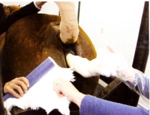
- Pack the rectum
Rectal packing involves inserting a tampon that extends from just proximal to the tear to the anus. The tampon is created by filling a stockinette with roll cotton. The tampon is well lubricated and inserted partially filled. Additional cotton can be inserted into the tampon after insertion. The tampon MUST extend the full length of the rectum from proximal to tear to anus; if it is not long enough, it will shift in position and actually create more issues as manure builds up in the tear.
Horses” AAEP Proc 2008
5. Hospitalization and monitoring
These horses should not be managed on the farm unless no other options exist. It is crucial to warn the referral center if you suspect or have identified a rectal tear. It is easy to make things worse if you are unaware of the situation.
Referral center treatment
Antibiotics and laxatives may be all that is required for grade I and II tears. These are combined with daily examination and careful emptying of the rectum as needed.
Some grade III tears will respond to conservative therapy including every 1-2h removal of feces, antibiotics, laxatives, packing, and epidural catheters to maintain atonic rectums. Healing takes approximately 2-3 weeks.
Surgical options exist but are not commonly pursued. These include placing a temporary indwelling rectal liner via ventral midline celiotomy, colostomy creation (temporary or permanent), and suture repair (via the rectum, via ventral incision or laparoscopically).
Prognosis
Complications include cellulitis, abscess formation, severe toxemia, peritonitis, laminitis, and recurrent intestinal obstruction from adhesions/strictures
- Grade I : 93-100% survival with conservative treatment
- Grade IIIa : 38-70% survival
- Grade IIIb : 38-69% survival
- Grade IV : rapidly fatal in most cases
Grade II are often diagnosed later during palpation. These often feel like divots in the rectum over each ovary. They do not cause issues unless they become impacted with feces.
Legal aspects
Rectal tears are a common cause of malpractice suits. Standard of care is important. Veterinarians need to show they took proper precautions and minimized straining by sedating the horse, using a twitch and using sufficient lubrication. If a tear occurs, the veterinarian should inform the owner immediately, assess the tear, start therapy and refer if indicated. The insurance agent should also be notified and can give advice on next steps.
Prevention
Minimize the risk of tears
- properly restrain and sedate the horse
- use sufficient lubrication
- do not try to force against straining or peristaltic wave
- do not try to palpate structures ahead of hand (go past and then palpate)
Owners should also be warned of the risks of rectal tears. This can be difficult to remember for each breeding evaluation or emergency colic. You may want to include a discussion of risks when beginning breeding work for the season and/or as part of your client education programming.
Resources
A review of equine rectal tears and current methods of treatment. 2015 Equine Vet Educ, Vol 27(4): 200-208
Evaluation of risk factors, management and outcome associated with rectal tears in horses: 99 cases (1985-2006). 2008 JAVMA 233(10):1605-1609
How to Perform First Aid on Rectal Tears in Horses. 2008 AAEP Proceedings; 64: 298-300- best resource for rectal tampons
Antimicrobial therapy for GI diseases in horses, 2003 VCNA, Vol.19(3), pp.645-663
