FA GI Topics
Small ruminant and Camelid GI disorders
Small ruminants
Small ruminants are not particularly predisposed to GI disorders except for bloat (eating too much fermentable feedstuffs). The most common surgery is a rumenotomy for gastric indiscretion. Small ruminants are not likely to stand for surgery so rumenotomy is typically performed with the animal in right lateral recumbency and under GA or heavy sedation. In most cases, the rumen will be distended and impairing respiration due to pressure on the diaphragm. The enlarged rumen will also make regurgitation very likely. Intubation allows better oxygenation, ventilation, more stable anesthesia management via inhalant anesthesia and minimizes the risk of aspiration if/when regurgitation occurs. Most small ruminants can be managed on standard surgery tables and with SA anesthesia machines.
Since GI disorders are uncommon, it is important to rule out urolithiasis as a cause of the abdominal distress.
Camelids
The most common cause of abdominal pain in camelids is uterine torsion. This is treated by rolling and/or abdominal surgery to detorse the uterus. The cria is usually close to term.
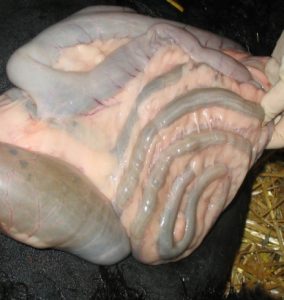
Camelids do occasionally need C1-otomies for gastric indiscretion or impactions. C3 ulcers present as mild colic but are not usually treated surgically. Camelids do form trichophytobezoars (hair+plant balls) and enteroliths that tend to lodge in the duodenum or spiral colon. GI tumors are also possible. Spiral colon obstruction is typically by enterolith. The spiral colon in camelids is tightly wound and more prone to obstruction than cattle. These camelids present with worsening signs of abdominal pain and decreasing manure production. Most camelids do NOT develop hypochloremic metabolic alkalosis. They do tend to become hypokalemic. Blood chloride values are not as useful as in cattle for identifying pyloric obstructions but C1 chloride levels have been found to be elevated with such lesions. Ultrasound guided abdominocentesis may also be useful. Barium studies can be useful for identifying obstructions in smaller animals.
Camelids do not typically show obvious colic signs. Abdominal exploratory should be considered with persistent, low-grade discomfort in a treated animal. Ultrasound is a key diagnostic tool for camelids. Digital rectal examination can be used to assess for feces; rectal palpation is often tricky and dangerous due to their smaller size. SI lesions will lead to more severe signs and more rapidly due to the normally high rate of secretions and fluid absorption.
Unlike cattle, the most common approach to GI obstructions or for GI exploratory is via a ventral approach under general anesthesia versus right paralumbar fossa in the standing animal. If a C1 lesion is expected, the incision is made in the left paralumbar fossa. For C2/C3, the incision is made either in the right paralumbar fossa or ventrally. .
Postoperative care often includes treatment with sucralfate and/or omeprazole to minimize the risk of gastric ulcer formation. Other complications include adhesions (between bowel loops) and stricture formation.
Other camelid disorders
Camelids are prone to megaesophagus and tooth root infections. Tooth root infections present as a draining tract or facial swelling and can sometimes be treated by prolonged antibiotic therapy but removal is often needed. Teeth are removed by oral extraction or by lateral buccotomy (bone flap).
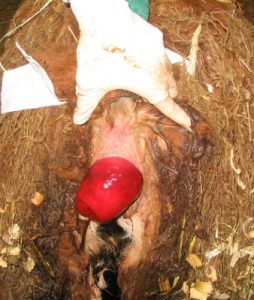
Rectal prolapses are not uncommon. These are most frequently associated with straining due to parturition, urolithiasis, coughing or parasites. Once the swelling is reduced using osmotic agents, most can be replaced and kept in position with a purse string suture. Resection may help minimize recurrence if needed.
Camelids may also have atresia ani. If it is a female, affected animals often have a rectovaginal fistula so they are able to pass manure. Owners may be unaware of the atresia. Males will develop severe obstipation at an early age.
Resources
Abdominal imaging in small ruminants: liver, spleen, gastrointestinal tract, and lymph nodes. VCNA FA 37 (2021): 55-74
Trichophytobezoar duodenal obstruction in New World camelids. Vet Surg 2005;34(5):524-9
Surgery of the gastrointestinal tract in camelids Part I AABP 2009; KD Newman, DE Anderson
blockage or partial blockage of the intestinal lumen by feedstuff
mineral concretion or calculus formed anywhere in the gastrointestinal system
the suture runs in and out in a circular pattern, so when pulled tight, it cinches off the center area like a purse string
