Equine Lameness
Palpation anatomy- forelimb
Find a horse and have fun!
by Vic Cox
The distal equine limb is mostly bone with some tendons, ligaments and neurovascular structures between the skin and bone. Most soft tissue structures can be palpated by pressing them against the bone. With tendons and ligaments you should press fairly hard but with vessels and nerves a softer touch will work best. The following instructions will serve as a guide but the list is not exhaustive.
Forelimb stay apparatus
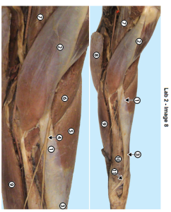
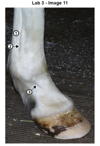
On the cranial side of the cubital (elbow) joint find a vertical cord that is hard when weight bearing and soft when weight is shifted off the respective forelimb. This tendon-like structure is the lacertus fibrosus = long tendon of the biceps m.. The lacertus fibrosus serves to connect the biceps and extensor carpi radialis tendons making a complete tendinous “cable” from the cannon up to the scapula. This forms the main part of the forelimb stay apparatus.
On the cranial edge of the lacertus fibrosus feel a cord like structure that can be moved on the underlying lacertus fibrosus. This is the cutaneous branch of the musculocutaneous nerve. It supplies cutaneous innervation to the medial side of the antebrachium and carpus.
Carpal joints and bones
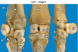
Pick up the limb and flex the carpus so you can palpate the radiocarpal and intercarpal joints on the dorsal aspect of the carpus. Note that both open wide when the joint is flexed and the spaces are partially covered over by the extensor carpi radialis and common digital extensor tendons (below). Carpal chip surgery is done between these tendons. Carpal chips, or a slab fracture in the case of C3, are found on the dorsal edge of the carpal bones.
On the palmar side of the carpus note the prominent “bump” caused by the accessory carpal bone (Ca). Wrap your fingers around the medial side of the carpus and palpate Ca with your thumb. Press hard above the Ca bone and feel the vertical “seam” between the two tendons that insert on Ca. Both these tendons have ulnar in their names (ulnaris lateralis and flexor carpi ulnaris). Between these tendons the ulnar n. is found and the dorsal branch of the ulnar n., a cutaneous branch emerges near the “seam” a few cm above Ca and runs laterally over the tendon of the ulnaris lateralis m.
Metacarpal tuberosity
Put the limb down and palpate the insertion of the extensor carpi radialis tendon on the metacarpal tuberosity on the upper dorsal part of the cannon bone just distal to the carpal bones. The tuberosity is a reinforcement of the bone to withstand the constant pull of the extensor carpi radialis tendon as part of the stay apparatus.
Extensor tendons
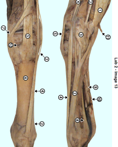
Grasp the cannon region firmly and rub your thumb over the dorsal aspect of the cannon bone. You should feel two tendons. Note that the smaller one is lateral and is the lateral digital extensor. The medial one is the common digital extensor tendon.
Splint bones
While still grasping the cannon region move lateral or medial and find the groove between the cannon and splint bones. Follow the splint bones distally to find the bulbous end of each splint bone which is sometimes referred to as a button. The splint bone is slightly longer on which side (M v. L)? Note that the distal part is flexible. It can be broken off on the lateral side by a kick from another horse.
Communicating ramus (nerve)
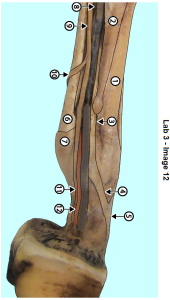
On the palmar surface of the cannon with a soft touch run your index finger up and down over the digital flexor tendons. You should find a cord like structure between the skin and the flexor tendons. This is the communicating nerve between the medial and lateral palmar nerves. Note the oblique course of this nerve, which side is it higher on?
Flexor tendons and suspensory ligament
On the medial and lateral sides of the cannon region move your thumb forward and backwards while grasping the cannon with your other fingers. From dorsal to palmar you should feel the cannon bone –> splint bone –> suspensory ligament (lower half of cannon only) –> digital flexor tendons.
Continue to grasp the cannon and now move your thumb up and down the lateral side of the flexor tendons and try to find a “seam” between the superficial and deep digital flexors. If you can’t, don’t worry about it.
Pick up the forelimb and grasp the flexor tendons between your thumb and index fingers. Slip the digital flexor tendons between your fingers and note that the DDFT is big and round in cross section while the SDFT is thin and flat.
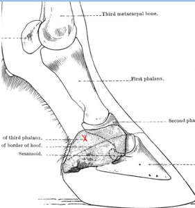
Put the limb down and find the suspensory ligament in the distal cannon using your thumb and index fingers to feel both medial and lateral sides. Note tension in the medial and lateral branches of the suspensory ligament which relaxes when the limb is not weight bearing. Follow the groove between the suspensory ligaments and the distal cannon down to the fetlock where it ends due to the strong horizontal ligaments that bind the sesamoid bones to the end of the cannon bone. The distal end of this groove is one site for injection into the fetlock joint.
Distal to the fetlock find the extensor branch of the suspensory ligament obliquely crossing the long P1. Remember that the main part of the suspensory ligament attaches to the abaxial aspect of medial and lateral sesamoid bones but some of the fibers form the extensor branch.
Palmar digital neurovascular bundle
Using your thumb and index finger grasp the sides of the fetlock joint and with a soft touch feel the bundle of digital vessels and nerves which are superficial here. The pulse is difficult to palpate here unless there is inflammation in the foot, especially in laminitis. It is good to routinely attempt to feel the pulse here so that when it is easily found you will have physical evidence of inflammation in the foot.
With the same two fingers slide down to the pastern region and feel the long pastern bone and the flexor tendons. Next find the groove between the bone and tendons. In this groove lies the palmar digital nerve. It is difficult to feel, but the groove between the pastern bone and the flexor tendons is a good landmark to block this nerve for suspected navicular disease. This is the most commonly blocked nerve in lameness exams.
Ergot
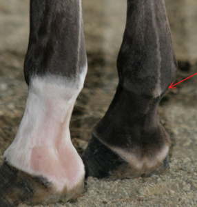
Palpate the palmar surface of the fetlock to find evidence of the ergot, a raise scaly structure. It also connects to the ligament of the ergot. This ligament is useless to the horse, but can be confused with the palmar digital nerve which is cut for navicular or heel pain. If you pull up on the ergot area with one hand while palpating the area described in #10 above lightly, you may feel the ligament of the ergot as a thin cord under the skin running distal and somewhat dorsally from the ergot region on both sides of the palmar pastern.
Collateral cartilages of the distal phalanx and digital cushion
Above the coronary band in the region of the quarters and heel feel the upper edge of the collateral cartilages of P3. “Wiggle” them and note that they are flexible. Put your thumb between them and push down on the digital cushion which fills the space between the collateral cartilages and is deep to the bulbs of the heel.
Resources
Horse anatomy A coloring atlas, Scribd
Anatomical Landmarks of the Canine and Equine Thoracic Limbs, UGA free ibook
The thoracic limb, UGA free ibook
