LA umbilical disorders
How to – Umbilical herniorrhaphy
Indications
Herniorrhaphy should be performed for smaller hernias where bowel entrapment is present or possible. It is commonly performed for most umbilical hernias and many incisional hernias. A firm hernial ring must be present prior to attempted repair. Developing a firm hernial ring can take 60 days after injury.
If external infection is present, this should be drained prior to surgery. Allow 5 days after drainage to allow granulation tissue to develop. To minimize infection, surgery should not be performed until granulation tissue is present.
Relevant anatomy
With a hernia, the linea alba is split, leaving a body wall defect in the center.
Preoperative management
Food restrictions: Withholding food makes hernia closure easier, particularly in ruminants. Food can be withheld for 48-72 hours in ruminating animals.
NSAIDs/analgesics: Preoperative NSAIDs are recommended.
Antibiotics: Antibiotics are given preoperatively if infection is likely (calves often have internal abscessation). Penicillin, ceftiofur or ampicillin are reasonable choices in food animals.
Tetanus prophylaxis is recommended in horses.
Local blocks: Inverted U shaped line block or TAP block
Position/preparation: The animal is positioned in dorsal recumbency. The ventral abdomen should be clipped fully to allow possible extension of the incision to the liver or to the bladder. The surgeon should be prepared for deeper exploration.
Surgery Supplies:
- Standard surgery pack
- #2 PDS or nylon for body wall closure
- 0 absorbable suture, taper needle, SQ
- 0 or 1 suture, cutting needle, skin
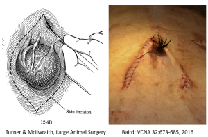
Surgical procedure
- A fusiform incision is made over the hernial ring, leaving enough skin for closure. Alternatively, an inverted V can be made around the prepuce in males with the prepuce reflected to expose the body wall
- Dissect loose connective tissue off the body wall.
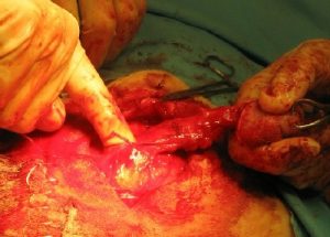
- Incise the ring on the lateral aspect (avoiding umbilical structures cranially and caudally), creating an incision into the peritoneal cavity large enough for a finger to enter.
- Palpate the ring as much as possible, identifying any stalks that extend into the abdomen through the ring (these are more likely infected structures).
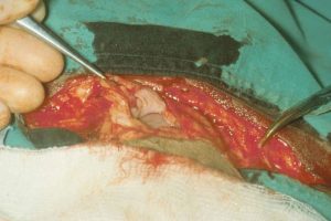
- Incise the body wall, following the ring on its inner aspect. Incise only those areas that are palpably normal (no infected stalks). Once the incision is large enough to visualize the umbilical structures, dissect around any stalk.
-
- In uninfected cases this will result in a circular opening into the peritoneal cavity and the hernia skin will be totally removed.
-
- If there is a stalk, continue circling the stalk until it is freed. Incise the skin to follow the stalk into the abdomen.
- If the infection continues to the bladder, remove the end of the bladder to remove the infection. Close the bladder in a two layer inverting pattern.
- If the infection continues to an umbilical artery, it will usually end in a bulb. Resect proximal to the bulb.
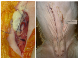
- If the infection continues to the liver, it may need to be marsupialized. For marsupialization, the infected vein is exited through a small incision made laterally. Trim off the stump and ligate the vein so it can be moved safely. Exit the ligated vein through the incision. The vein is tacked to the skin. The infection will drain when the animal is standing. The stump can be amputated the following day. Avoid flushing for at least two days, even if drainage is limited. Note: these animals are high risk anesthesia patients and will usually develop a hernia at the marsupialization site.
- If there is a stalk, continue circling the stalk until it is freed. Incise the skin to follow the stalk into the abdomen.
- Closure
- Close the body wall with #2 PDS or nylon, simple continuous pattern
- Close the subcutaneous tissues with 0 absorbable suture, simple continuous pattern
- Close the skin with 0 or 1 nonabsorbable suture, Ford Interlocking pattern
Postoperative care
- Stall rest with handwalking for 60 days (adult) or at least 3 weeks (young animal) to permit body wall healing
- Continue antibiotics if any contamination
- Continue NSAIDs 2-3 days
- Refeed slowly
Complications
- Peritonitis- fever, depression, poor motility
- Hernia reformation
- Incisional infection or dehiscence– signs of inflammation or intestines protruding from the incision
Videos
- Simple hernia: youtube link
- Approach to a more complex hernia: youtube link. Not sure how the banana part got in there, TBH.
Resources
AN Baird. Surgery of the umbilicus and related structures. VCNA 32:673-685, 2016
suture pattern in which tissue edges are turned inward
sutured edges come apart; lack of healing of incision
