Food Animal Male Urogenital Surgery
How to – Epididymectomy
Indications
Bilateral epididymectomy is performed to create a non-fertile male that can still identify animals in heat and/or retain male secondary sex characteristics (lion mane, etc). Unilateral castration is performed for unilateral disorders of the epididymis that require removal.
Animals selected as teasers should be manageable, of a size to not cause injury during mounting behavior and have good libido.
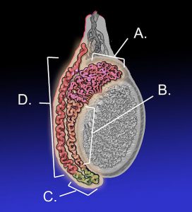
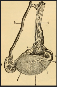
Relevant anatomy
Preoperative management
Food restrictions: In bulls, the procedure can be done standing in a chute with no sedation. In most other cases, the animal is only lightly sedated for the procedure and no food withholding is required. If needed for exotic ruminants or other cases, food should be withheld for 48 hours to minimize bloat.
NSAIDs/analgesics: Preoperative NSAID administration is recommended to minimize pain and inflammation. Postoperative NSAID administration is not usually required unless inflammation or pain is evident.
Antibiotics: Preoperative antibiotics are not indicated for routine cases.
Tetanus prophylaxis is recommended.
Local blocks: Lidocaine is used to block the cord and/or the surgical site. A cord block avoids distortion of the surgical site. A ring block around the cord can help with analgesia.Injection along the proposed incision line and into the epididymis is quick and effective but does alter the anatomy. 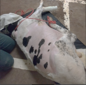
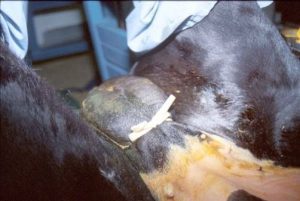
Position/preparation: Bulls can be restrained a chute with a butt bar in place. Sheep are held in a sitting position. Other animals are cast or sedated until in lateral recumbency. The upper leg should be pulled forward to expose the scrotum. The scrotum is clipped (if needed) and scrubbed prior to and after the local block. Surgeons should glove. A headlamp is recommended.
Surgery Supplies:
- Scalpel blade + handle
- Allis tissue forceps or towel clamps
- Needle holders
- Suture scissors
- 3-0 to 0 absorbable suture depending upon patient size
Surgical procedure
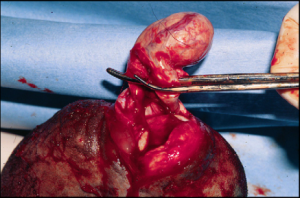
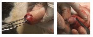
A clean towel or sterile drape can be placed under the scrotum and/or over the animal’s abdomen for suture management. Stabilize the testicles in the scrotum with your nondominant hand or with a penrose drain wrapped around the scrotum to hold the testicles in place. The tail of the epididymis should be clearly visible and the testicle should not be able to move in the scrotum.
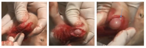
Incise over the tail of the epididymis, exposing both the site at which it is connected to the testicle and the connection to the narrower ductus deferens. Dissect through the skin until you can see the epididymal bulge. Expand the incision so you can see where the epididymis ends and the testicle is visible.
Option 1. Ligate and then dissect
Continue your incision through the vaginal tunic, exposing the epididymis. One side will connect to the ductus and has a relatively narrow area to ligate. The other side connects to the testicle and is going to be harder to define where best to place your suture. It doesn’t matter. The discontinuity you are creating is the important part. Find one side and ligate it. Repeat on the other side. Dissect the epididymis free from the testicle and remove it. Close either the tunic or the skin using 3-0 or smaller absorbable suture in a continuous pattern.
Option 2. Dissect and then ligate
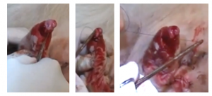
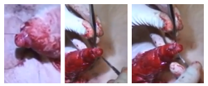
Grasp the epididymis (still covered by the vaginal tunic) with the Allis tissue forceps. Dissect the epididymis off at the junction with the testicle. Continue until you have the epididymis only connected to the other structures by the ductus deferens. Ligate the ductus with 3-0 absorbable suture. Cut the epididymis off. Close the tunic is a simple continuous pattern using the 3-0 absorbable suture. Leave the skin open.
Postoperative care
- Monitor for signs of infection. Open for drainage if necessary.
- Avoid breeding or access to females for 8 weeks.
- Communicate meat withholding restrictions
Complications
There is a risk of hemorrhage and damage to the testicle; resulting testicular atrophy could alter secondary sex characteristics. There is a risk of continued fertility, primarily due to recanalization of the ductus. Sperm granulomas likely occur but have minimal impact on health of the teaser animal.
Videos
Testicular block
Cord block
Scrotal ring block
Dissect first approach -video
Closed approach– video
Dr. Knauers demo of dissection
Resources
Preparation of teaser bulls – Veteriankey
Epididymectomy on a cadaver (starts at 5:50)- good view of anatomy
