Bovine musculoskeletal disorders
Neonatal musculoskeletal injuries
These injuries are associated with the birth process, often with dystocias.
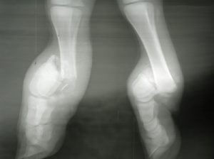
Cannon bone fractures
These are the most common injury associated with dystocias and human interference.
The fracture tends to involve the distal physis of cannon bone. These are usually closed, simple and relatively stable if alignment can be achieved (the physis interlocks to hold the bones in place). The fractures are sometimes accompanied by vascular damage. Vascular damage will show up later and can delay or prevent healing. Calves may not have stood to nurse or absorbed colostrum well, so may need colostrum or plasma.
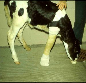
Internal fixation does not work in calves as their bone is too soft. Treat cannon bone fractures with a cast or Schroeder-Thomas splint.
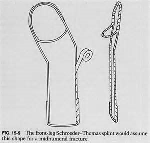
A full limb cast is ideal to stabilize joint above and below the fracture; however, with lower limb fractures, often a half limb cast is sufficient and is much easier for the calf to manage.
Prognosis is guarded due to the potential for blood supply damage. Blood supply damage may not be evident until the cast change at 2-3 weeks. Most do fine but it is hard to know in advance.
Cast cutters can cut skin! Especially in large animal species with minimal soft tissue coverage. If you create a wound with the cast saw, do not close it (fiberglass particles in the wound). Just clean it up.
Rib fractures
Rib fractures are related to posterior/breech presentation. Careful palpation is necessary to diagnose these. The fracture ends can rupture thoracic vessels. Treatment involves restricted exercise and/or stabilization. Flail chests require stabilization.
Jaw fractures
The jaw can be damaged by jaw pullers used for dystocia. Check for colostrum absorption (the calf may not have nursed). These have a variety of stabilization options but need something.
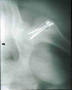
Slipped capital femoral epiphysis
The physis is weaker than the round ligament of the femur. Instead of hip luxation, the physis breaks. The hip stays in the socket, femur gets loose. These occur with hip lock dystocia – often in double muscled breeds. Diagnosis may be delayed. Radiographs are useful for diagnosis.
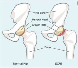
Prognosis is fair with surgery, poor otherwise. FHOs (femoral head ostectomy) are not a good option in cattle.
Femoral fractures are somewhat stabilized by the muscles of the upper limb and can heal with conservative management. However, cattle lie with the limb abducted and these forces will likely lead to abnormal limb angulation. There is s report of fixing a femoral fracture with external skeletal fixation, as well.
Femoral nerve paresis “patellar luxation”
These are not truly patellar luxation but the quadriceps is not working. These are congenital lesions and often bilateral lateral luxations. The problem is with the femoral nerve. And, as the patellar doesn’t move correctly the groove doesn’t form properly. Calves will have a crouched stance. Poor prognosis if no return of nerve function.
Synovial infections
Synovial infections are typically associated with umbilical infections, respiratory infections or GI infections. One or more of the large joints may be affected. Prognosis is guarded and the cause of the infection needs to be addressed (umbilical resection, etc). See joint issues chapter.
