Swine, SRC and poultry lameness
Camelid orthopedics
Anatomy
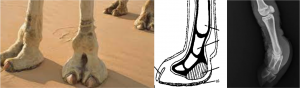
Camelids bear weight on both P2 and P3 rather than just on P3 as with most large animal species. There is no navicular bone. They walk on double pads with a claw in front. The claw is nonweightbearing but is important for traction and propulsion. A digital cushion supports both P2 and P3.
Other notable anatomical features:
- In most animals, the medial and lateral compartments of the fetlock joint are separate (unlike in cattle).
- The superficial digital flexor tendon has a direct fascial connection to the proximal suspensory ligament (so issues with one tend to impact both)
- More in Chapter 58 – Musculoskeletal Surgery of Llama and Alpaca Care
- Smaller camelids can manage with 3 limbs so amputation is a feasible option. Similarly, postoperative care and fracture management are easier due to the ability to use slings and various splints. A light weight body also means camelids can be treated with implants designed for dogs, horses or humans; hence, a wider variety of options exists for repair of injuries or abnormalities.
Foot issues
Camelids are not prone to foot rot but can get interdigital dermatitis if conditions are right (moisture + trauma). The same conditions and organisms can cause ulcerative pododermatitis of the foot pads. Treatment requires removal of any loose skin or pad, antiseptics and protection with a wrap or bootie. The animal should be kept in a clean, dry environment. Antibiotics may be needed for deep infections.
The dorsal interdigital region and heel area are both common sites for chorioptic mange.
Limb deformities
Limb deformities can be angular (in the lateral plane) or flexural (in the dorsal-palmar plane).
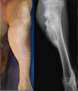
Mild bilateral carpus valgus is prevalent in camelids. More severe angular limb deformities may be related to hypophosphatemic rickets. Crias with dark hair coats are especially prone to developing low vitamin D levels because of reduced absorption of sunlight by the skin. Crias born during the cloudy months should be supplemented with 1000 IU/kg vitamin D3 subcutaneously (SQ) every 60 days. Physitis due to rapid growth has also been identified. Weaning the cria may be needed to slow the rapid growth and allow the physis to recover.
Animals with physitis typically have one side that grows faster than the other side; this leads to the angular deformity. Camelids with severe angular limb deformities can be treated with surgery to slow the growth of the faster growing aspect. We do this with a screw placed across the physis (transphyseal screw) or by bridging the physis with a figure 8 wire that prevents growth, similar to that done in horses [see equine chapter]. Camelid growth plates close much later than horses so more time is available for correction.
Flexural deformities are sometimes encountered and are treated similarly to foals. In these cases, the tendons are “tight” and restrict the limb motion so that it can’t straighten. In camelids, we can be more aggressive with surgery due to the limited athletic requirements.
Suspensory degeneration /Fetlock hyperextension
Camelids can develop “dropped fetlocks” or bilateral conformational abnormalities due to what is believed to be suspensory degeneration. This syndrome has been associated with mineral deficiencies, abnormal proteoglycan levels in the suspensory ligament and genetic predisposition. We see similar lesions in horses. In both species, no effective therapy exists and prognosis is poor.
Unilateral cases of dropped fetlocks can occur with excessive weight bearing and are more likely to be due to suspensory damage rather than degeneration.
Juvenile osseous sequestration
While horses and cattle develop bony sequestration after wounding, many of the sequestra diagnosed in camelids do not have open wounds associated. This form of sequestration is most common in young camelids and it more closely resembles a syndrome seen in children in which bacterial emboli seed bone, leading to bone ischemia.
Long bones of the appendicular skeleton seem to be most affected. Animals present with signs of heat, pain, local swelling and lameness. Diagnosis is made radiographically.
Sequestrectomy (removal of the sequestrum) usually results in a good prognosis as long as enough supporting bone remains to prevent fracture. Culturing the bone is recommended
Joint issues
Stifle ligamentous injuries (cruciate tears) are not common but can be found in camelids. Both intracapsular and extracapsular repair approaches have been successful.
Patellar ligament tears have been reported in pack llamas and in breeding males. Affected camelids walk with a crouched posture (stifle stays flexed) and are unwilling to bear weight fully. Diagnosis is confirmed via surgery. Various forms of repair have been successful short term, but reinjury rates are high.
Shoulder luxation is uncommon but is seen in young male alpacas. Surgical repair is generally successful. Animals are kept in a Velpeau sling in the early postoperative period.
Hip luxation is treated as it would be in dogs.
General arthritic pain is treated with meloxicam and supportive care.
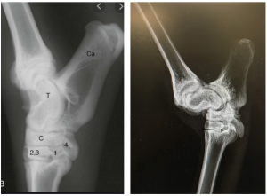
Patella luxation
Patellar luxation can be due to trauma or to congenital conformational abnormalities. Animals castrated early in life (<12 mo old) tend to develop straight hindlimbs and are considered more prone to patellar luxation. Treatment involves lateral imbrication and release (surgery to tighten up the ligaments on one side while releasing the ligaments on the other side). Prognosis may improve if the surgery is combined with trochleoplasty (reconstruction of a deeper groove in the femur so the patella is less likely to slip out of position). Recurrence of the luxation after surgery is common. A sling may be needed for the first two weeks postoperatively unless trochleoplasty is performed.
Septic arthritis
Septic arthritis occurs primarily due to trauma (direct wound into the joint). Hematogenous septic arthritis is less common than in cattle and horses. Diagnosis of infection includes joint cytology and culture. Treatment is similar to other species and is focused on joint lavage and antibiotic therapy (iv antibiotics, intra-articular antibiotics, and/or iv regional perfusion of antibiotics).
Congenital lesions
Camelids have significant numbers of congenital lesions. Orthopedic lesions include wry nose, polydactyly (extra toes), syndactyly (fused toes), patellar luxation, flexural deformities and rotated talus bones. The rotated talus bones are associated with an abnormal gait.
Key Takeaways
Camelids do not get too many lameness issues.
Be aware of
- suspensory degeneration leading to fetlock hyperextension
- juvenile osseous sequestration unrelated to open wounds
- patellar luxations, potentially related to early castration
Camelids can be treated much more like dogs in terms of joint luxations, etc. Their light bodies mean they are able to ambulate on three limbs and we have more repair options.
Resources
Pain management in small ruminants and camelids: Analgesic agents.Vet Clin North Am Food Anim Pract . 2021 Mar;37(1):1-16.
Llama and Alpaca Care. Medicine, Surgery, Reproduction, Nutrition, and Herd Health. Elsevier, 2014.
limb bows outward at the carpus so th
inflammation of the growth plate or physis
pieces of dead bone that cause persistent draining tracts as the body tries to liquefy them
