FA GI Topics
How to – Rumenostomy
Indications
Rumenostomy is performed in growing calves with chronic bloat and in any age ruminant to provide nutritional support. Rumenostomies can be performed instead of rumen trocarization for improved results. Rumenostomies are also performed for feed trials to enable direct feeding and/or sampling. Finally, a rumenostomy can be placed in an adult cow to provide easy access to microflora (rumen donor). Rumenostomies typically have smaller openings designed to close over time (calves) or are kept open with a rumen cannula sized to fit the need.
Relevant anatomy
The rumen is located on the left side of the abdomen. A rumenostomy is centered in the left paralumbar fossa.
Preoperative management
Food restrictions:
Surgery is harder with a full rumen. Animals should be held off feed for 24 hours if not urgent. Bloating calves are NOT typically held off feed as the procedure may be needed on an emergency basis.
NSAIDs/analgesics:
Animals should be given flunixin meglumine or other NSAID prior to surgery
Antibiotics:
Procaine penicillin G is typically given preoperatively
Local blocks:
Line block, inverted L or paravertebral block with lidocaine
Position/preparation:
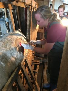 Standing and restrained. Larger animals can be restrained in chutes or headgates; younger animals are often restrained by people.
Surgery Supplies:
- Standard pack
- Scalpel blade and handle
- 2-0 to 0 absorbable, taper needle for the inner layer
- 2-0 to 0 nonabsorbable or absorbable, cutting needle for the outer layer
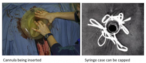
- Cannula (optional); soaking in hot water
- Syringe case, 0 suture, umbilical tape (optional)
Surgical procedure
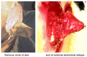
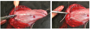
After the area is clipped, prepped and blocked, a circular incision is made in the center of the flank through the skin. In a standard rumenostomy, this is the diameter of two ribs. If a cannula is being inserted, this is the inner diameter of the cannula. The circle of skin is removed. A circle of external abdominal oblique muscle is removed.
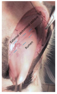
The internal abdominal oblique and transversus muscles are separated along their fibers to grid the opening. This creates a natural valve effect that minimizes rumen fluid leakage.
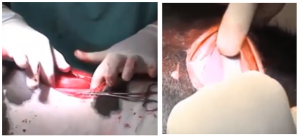
The rumen is grasped with allis tissue forceps and pulled gently out the incision until is “poofed out” and is filling the the hole. Some exteriorization is needed to minimize tension on the suture line. Excessive exteriorization creates folds and areas that can necrose under a cannula.
The rumen is secured to the external abdominal oblique with an inverting pattern using 2-0 or 0 absorbable suture (taper needle). This pattern (blue lines in image) is placed as far from the center of the circle as possible to avoid interference with the final layer. This pattern creates a water tight seal that protects the abdominal cavity from contamination.
A circle of rumen is removed stepwise. One quarter of the arc is incised. The cut edge of the rumen is sutured to the cut skin edge in a continuous appositional pattern using the 2-0 or 0 suture (cutting needle).
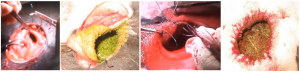
After the first quarter is incised, another quarter is transected and sutured. At this stage, the continuous pattern should be knotted to prevent a purse string effect. The suture is not cut free but is simply tied in place and then continued in use. The third quarter is transected and sutured. This is continued until a full circle of rumen wall is removed and the remaining rumen secured to the skin.
If a cannula is being placed, it is inserted at this time. Having it soaking in hot water helps the pliability. It should fit snugly. A temporary cannula can be constructed out of a syringe case for cases of bloat. The case is secured to loops of suture using umbilical tape. The cap can be put back on the case to minimize fluid leakage; bloat will blow the cap off and the gas can escape. Both can keep the site open, potentially longer than needed.
Postoperative care
- NSAIDs are continued for 2-3 days, antibiotics for 5 days
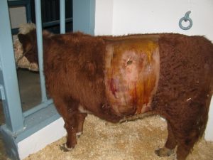
- The flank is coated with vaseline and cleaned as needed to minimize scalding from the rumen juices.
- Nonabsorbable sutures are removed at 10-14 days
- Without a cannula, the rumenostomy site will gradually shrink and eventually close in most situations. If sized appropriately, the bloat should be resolved by the time it closes.
Complications
Peritonitis- animals may have a fever and be off feed. Some cattle can wall off the infection.
Incisional infection- part of the suture line may need to be opened to allow drainage.
Skin scald – can be treated with cleaning, zinc oxide and vaseline
Lack of closure – with constant manipulation from cleaning or continued cannulation, the site will stay open. It can be closed if needed. Closure involves resecting the fistula and should be done by an experienced surgeon.
Videos
These videos were taken as part of a teaching laboratory; calves are in lateral recumbency rather than standing:
gridding for rumenostomy – close up video
Resources
a hole made into the rumen designed to stay open for awhile
suture pattern in which tissue edges are turned inward
tissue layers are lined up in apposition; direct approximation of layers
