Equine podiatry
Foot anatomy -imaging
Advanced imaging of the foot, Vic Cox
A study of foot bandsaw sections is helpful for understanding CT and MR images. CT is best for mineralized tissues (like conventional radiographs) while MRI is best for soft tissue. MR images are brightest for tissues containing water or fat because MRI depends upon signals from the protons of hydrogen atoms and these are most commonly found in water and fat. For that reason, cortical bone will be dark while medullary bone will be bright (as seen below). Likewise, synovial fluid will be bright and inflammation (edema) will be brighter than normal tissue in MRI images
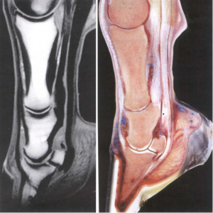
Note the bright medullary bone and dark cortical bone in the MR image on the left. Also note how synovial fluid is bright and articular cartilage is dark. Likewise, the DDFT and the straight distal sesamoidean ligament are dark. *=fibrocartilaginous part of DDFT on right.
Reminder of section types:
- Sagittal and parasagittal sections. A sagittal section divides the foot into nearly symmetrical medial and lateral halves. Any parallel plane is called a parasagittal section.
- Transverse sections are perpendicular to the long axis of the distal limb.
- Frontal (dorsal) sections are perpendicular to both sagittal and transverse planes.
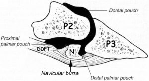 This is a sagittal section showing the DIP (distal interphalangeal joint) with three distended pouches. Not labelled is the thin distal sesamoidean impar (unpaired) ligament that binds the navicular bone to P3. Because the DDFT curves around the navicular bone, this bone acts as a trochlea (pulley); hence the term podotrochlear apparatus (bone and ligaments).
.
The impar ligament attaches to the distal edge of the navicular bone while the paired collateral sesamoidean ligaments attach to the proximal edge of the navicular bone. The sesamoidean ligaments are collateral ligaments because there are medial and lateral parts that wrap around the pastern bones and attach to the distal part of P1 and also to the “sides” of P2. The collateral sesamoidean ligaments are also known as the suspensory ligaments of the navicular bone. These ligaments counter the force of the distal end of P2 which pushes down on the navicular bone when the foot makes contact with the ground.
Note that as the DDFT passes distally it gets wider but thinner. The wide thin part is prone to tearing, but the lesion is difficult to see because the DDFT is dark on MRI although tearing may result in enough inflammation or granulation tissue to suggest the injury. Proximal to the navicular bone the DDFT is thicker and has a fibrocartilaginous part (asterisk in fig. 1) but best seen on transverse sections. In the region of the navicular bone the DDFT is divided by a groove into medial and lateral parts.
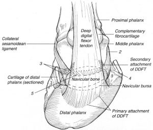
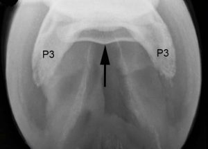 Dorsosolar radiograph of the navicular bone showing the axial ridge (arrow) on the flexor surface of the navicular bone. The palmar parts of P3 are sometimes referred to as the “wings” of the coffin bone. This radiograph reveals good differentiation between the cortical and medullary parts of the navicular bone. Note that the navicular bone is deep to the apex of the frog. Also note that the wall is more radiodense than the sole and the frog looks like the letter “A”.
Resources
Imaging the Equine Foot. Vet Clin Equine 37 (2021) 563–579
