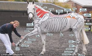
Equine bone disorders
Neck, back and pelvic pain
Osteoarthritis, osteochondrosis and muscle disorders can affect the neck, back and pelvic region. These often require referral for advanced diagnostics due to the difficulty in imaging these areas.
Neck pain
Disorders in the neck region include nuchal bursitis, osteochondrosis (wobblers), and osteoarthritis. Fractures of the neck are rare but are difficult to manage. Horses with neck pain often present with stiffness, reluctance to turn the head to the side, unwillingness to work on the bit or to eat off the ground. Stumbling or ataxia in the hindlimbs may be apparent due to compression of the spinal cord.
Physical exam findings may include focal sweating as well as signs of inflammation (most common with nuchal bursitis). Neurological abnormalities and limb lameness should be excluded.
Nuchal bursitis
Nuchal bursitis is not common but the region should be checked for heat, pain and swelling. Horses are often unwilling to flex the neck. Ultrasound is very useful for diagnosis. Fluid should be collected for cytology and culture. Endoscopic debridement and lavage is combine with NSAIDs for treatment. Antimicrobials are used in cases of septic bursitis. Medical treatment alone often results in recurrence of the bursitis.
Intervertebral facet joint OA
Neck OA can lead to unwillingness to bend the neck, difficulties with collection and/or jumping, and/or neurologic signs in the hindlimb. Diagnosis can be confirmed using radiographs, ultrasound or scintigraphy. Treatment includes oral NSAIDs and/or facet injections with corticosteroids and hyaluronic acid.
Back pain
Back problems are often secondary to hock issues but can be primary. Primary issues include saddle fit issues, overriding or impinging spinous processes, and muscle disorders. Due to the heavy muscle coverage, diagnosis and treatment can be challenging. Pain on palpation if usually evident, with horses reluctant to flex their back. Radiographs, ultrasound and scintigraphy are useful for identifying lesions; local anesthesia is needed to confirm lesions as sources of pain.
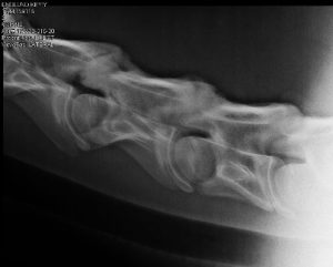
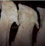
Overriding dorsal spinous processes
“Kissing spines” are a commonly reported cause of back pain. This disorder can be associated with poor saddle fit, poor conformation, poor conditioning, and rider/horse mismatch in size. Riders may report vague poor performance, change in head carriage, and/or unwillingness to bend. The thoracic area (and occasionally lumbar area) is painful on palpation. Imaging (radiographs, scintigraphy) alone is NOT sufficient to diagnose pain due to kissing spines. Abnormalities are common. Local anesthesia is needed to confirm the cause of clinical signs.
Treatment can be medical/conservative or surgical. Medical treatment involves local injections of steroids, physiotherapy and/or extracorporeal shockwave therapy. Repeated treatment every 6-12 months is to be expected. Standing surgery to either cut the interspinous ligaments or to remove every other affected dorsal spinous process is recommended for horses that do not respond to medical therapy or those with severe changes. Postoperative rehabilitation (strengthening, stretching) is important.
Suprasinous desmitis
Suprapinous desmitis is a cause of sudden back pain. Ultrasound is used for diagnosis. The area between T15 and L3 is most commonly affected. Note that horses may have ultrasonographic abnormalities that are not associated with back pain.
Intervertebral facet joint OA
Similar to the neck, the vertebrae in the thoracolumbar spine can be arthritic. Radiographs, ultrasound and/or scintigraphy can be useful for diagnosis but must be combined with local anesthesia or other signs that the area is causing pain. Treatment is via injections of steroids and hyaluronan, systemic medications and exercise.
Pelvic pain
Sacroiliac disorders
Disorders of the sacroiliac region cause poor propulsion and mild hindlimb lameness. These disorders are usually chronic. The horse may be painful on palpation of the tuber sacrale and positive on upper limb flexion. Owners may describe the horse as bunny hopping at the canter. For confirmation of diagnosis, other causes of hindlimb pain need to be excluded. Ultrasound (including transrectal ultrasound) and scintigraphy are useful to identify change in the region. Diagnostic analgesia is risky as it can impact the sciatic nerve, leading to severe hindlimb weakness or inability to bear weight on the limb. These can cause extreme stress in the horse and result in secondary injuries. Treatment includes peri-articular injections of steroids and treatment of any desmitis with intralesional injections of stem cells or similar products and shockwave.
Coxofemoral joint
Rare. Significant lameness with gluteal atrophy is often present unless the cause is OA or damage to the round ligament of the femur. Diagnostic analgesia is combined with imaging findings (scintigraphy, ultrasound, radiography). The hip joint can be injected using ultrasound guidance.
Muscle disorders
Muscle tears, strains and cramps can be severely painful. Minor changes in muscle function/tone will most often lead to poor performance, with decreased propulsion, coordination and stamina. Exertional myopathies such as polysaccharide storage myopathies and recurrent exertional rhabdomyolysis will present in this manner and are associated with elevated muscle enzymes. Muscle atrophy will obviously cause poor propulsion and can be caused by a variety of neurologic and myologic disorders, including equine protozoal myelitis, vitamin E deficiency, trauma and immune disorders. Careful physical examination is needed to determine if any atrophy is focal or generalized. Diagnostics include laboratory tests for muscle enzymes and vitamin levels, muscle biopsies, genetic testing and imaging tests. Therapy is specific for the disorder identified.
Resources
Neck, Back, and Pelvic Pain in Sport Horses, Vet Clin Equine 34 (2018) 235–251 – nice summaries, resources and images
Muscle Conditions Affecting Sport Horses. Vet Clin Equine 34 (2018) 253–276 – thorough review by a UMN researcher
Long-term prognosis for return to athletic function after interspinous ligament desmotomy for treatment of impinging and overriding dorsal spinous processes in horses: 71 cases (2012–2017). Veterinary Surgery. 2019;48:1278–1286.
Diagnosis, treatment and outcome of cranial nuchal bursitis in 30 horses. Equine Veterinary Journal 50 (2018) 465–469
How to evaluate a saddle as a cause of back pain. AAEP Proc Vol 62, pp 301-305, 2016. Try English saddle fitting for OTTBs and https://www.facebook.com/TheHorse/videos/10154446097148283/ to access the material if you can’t find the resource.
