Female urogenital surgery
How to – 3rd degree rectovaginal tear repair
Indications
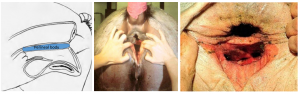
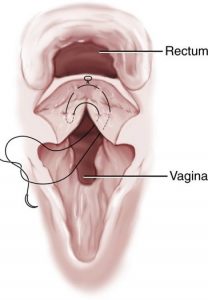
3rd degree tears are the result of dystocia. The baby splits the roof the vagina, the perineal body and the floor of the rectum. This creates two tubes that do not extend the normal distance.
Relevant anatomy
The trauma creates two tubes that do not extend the normal distance. Neither tube has a sphincter (no anal sphincter, no vulvar lips).
Preoperative management
Food restrictions: Mares should be on a low bulk diet – mostly grass and easily digestible feedstuffs. No hay. No dietary change is needed for most cattle due to the softer feces.
NSAIDs/analgesics: Perioperative NSAIDs are recommended.
Antibiotics: Perioperative antibiotics are needed.
Tetanus prophylaxis is recommended for horses
Local blocks: Epidural
Position/preparation: This is an advanced procedure; the surgeon should have advanced skills and a hospital environment is best. The procedure is done at least 3-4 weeks after the injury to allow fibrous tissue formation. The patient is standing and the surgeon is gloved.
Surgery Supplies:
- Standard surgery pack
- Headlamp
- Retractor suture or speculum
- 3-0 absorbable suture, taper needle
- 2-0 absorbable suture, taper needle
- 2-0 suture, cutting needle (Caslicks)
- headlamp
Surgical procedure
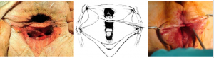
The goal of the surgery is to recreate the anal tube, the vaginal tube, the perineal body and some version of sphincter for each.
Create anal tube and vaginal tube
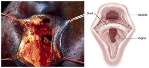
The shelf between the rectum and vagina is split horizontally, keeping the rectal side thicker than the vaginal side.
The incision is continued along both walls to the external mucocutanous junction. The wall dissections are undermined up the wall of the rectum and down the wall of the vagina to create two shelves that easily pass the midline.
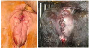
Once the shelves are created, they are apposed in three layers – the floor of the rectum, the perineal body, and the roof of the vagina. This can be done in a 6 bite technique all at once or in steps individually.
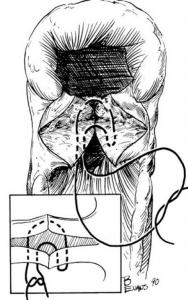
For the individual layers, 2 or 3 suture lines are run in tandem. One line closes the rectal floor using a simple continuous pattern. The perineal body and vaginal roof can be closed individually or in combination. A few bites are taken with one strand of suture, then a few bites with the other. The strands are alternated until the tube reach the level of the perineal body, approximately 1-2″ from the skin.
Once that level is reached, it is followed by perineal body reconstruction and a Caslicks.
Postoperative care
- Soft feces are maintained until recheck. The incision lines can be checked in 7-10 days for any fistulas needing further repair.
Complications
Fistula formation is common, particularly with insufficient undermining of shelves and firmer feces.
Videos
Resources
Vulva, vestibule, vagina and cervix, Equine Surgery
