LA Respiratory Issues
Nasal Discharge
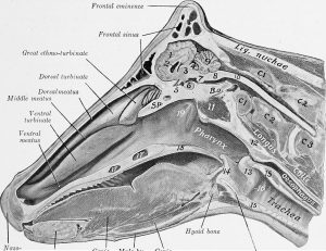
Anatomy Review
Nasal discharge anatomy powerpoint
Nasal discharge anatomy narrated powerpoint
Due to the complete nasal septum in horses, most unilateral discharge can be localized to the ipsilateral pharynx, sinus and paranasal region. Even though discharge from one guttural pouch opening or the ethmoid region could flow out the opposite side, it usually doesn’t. Tracheal and pulmonary fluids do flow out both nostrils.
Exudates typically present in 3 flavors: serous (normal), hemorrhagic or purulent. A combination of hemorrhagic and purulent discharge can be seen with ethmoid hematomas, neoplasia and occasionally with strangles (Strep equi). Lack of airflow can indicate congenital lesions or acquired obstructions. The most common congenital cause of no airflow is choanal atresia (lack of nasal passageway development; next chapter).
Often acquired obstructions are due to neoplasia or conchal cysts. Conchal cysts are congenital lesions that develop within the nasal turbinates; these grow slowly over time and can totally occlude the nasal passage.
Diagnostics include oral examination (looking for dental issues, particularly diastemata or gaps between teeth), radiographs ( can be very difficult to interpret), endoscopy, and sinuscopy (scope via the sinus; this usually requires sinus lavage first). More recently, standing CT has become more available and is very useful for diagnostic evaluation.
Challenge Yourself
Level A
