FA GI Topics
Abomasal displacement
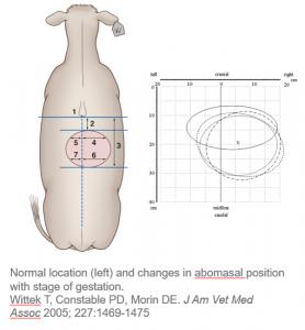
Most abomasal displacements are left sided and occur within the first 2 months after calving in grain fed dairy cattle, particularly Holsteins. During gestation, the abomasum often shifts from just to the right of midline to a horizontal position across the cranial abdomen. When it becomes gas filled in this position, the abomasum can now displace to either the left or the right sides of the abdomen. The pylorus will stay more ventral while the body of the abomasum floats more freely upward.
Right displaced abomasum
Right sided abomasal displacements can occur in any animal with an abomasum; however, they are rare in small ruminants. Beef and dairy cows are at equal risk and all ages can be affected.
Left displaced abomasum
Classically, left displacements of the abomasum occur in the early (first 4-8 weeks) postpartum dairy cow.
LDA Pathogenesis
- Decreased abomasal motility or increased gas production can lead to gas build up within the abomasum which allows for dorsal (floating) movement of the abomasum.
- During gestation, the uterus pushes up on the rumen, decreasing its ability to expand. After parturition, there is more room in the abdomen and less rumen fill, increasing the risk of an LDA. Further, post-partum cows have an increased risk of:
- GI atony due to hypocalcemia, diet change, or other diseases
- gas production due to altered fermentation with dietary changes.
LDA Risk Factors
- Man-made disease!
- Feeding increased concentrates and decreased fiber – this means cows don’t have a thick fibrous mat in the rumen
- Increased grinding of feed – this increases VFA production (good for milk production) and also increases microbial gas production
- Decrease rumen fill (post-partum)
- Concurrent diseases post-partum – coliform enteritis, mastitis, milk fever, metritis, retained placenta. These typically lead to ketosis, hypocalcemia and/or abomasal ulcers
Clinical findings
- scant loose feces
- ping on a line from tuber coxae to elbow, centered on 12th ICS
- sunken PLF (paralumbar fossa) with LDAs
- rumen usually pushes the left body wall out. With an LDA, the abomasum pushes the rumen toward the center line
- concurrent diseases (see above): ketosis, hypocalcemia and abomasal ulcers
- Sequestration of fluid within the abomasum and concurrent anorexia:
- dehydration
- metabolic alkalosis
- hyponatremia, hypochloremia, and hypokalemia
- paradoxical aciduria (Challenge: What is the pathogenesis behind this? Review here)
- Metabolic Profile and Inflammatory Responses in Dairy Cows with LDA, Animals (Basel). 2015 Dec; 5(4): 1021–1033.
- Ping location can help differentiate DAs from other GI disorders. An RDA cannot be easily differentiated from an abomasal volvulus or duodenal obstruction. As an abomasal volvulus requires emergency surgery, never let the sun set on a right sided ping!
Low serum chloride generally indicates a gastric outflow obstruction. The gastric HCl is normally reabsorbed in the duodenum. In cattle, gastric outflow obstruction is more commonly seen with DAs or abomasal volvulus but it can occur with pyloric lymphosarcoma or very proximal duodenal strictures (eg from a misplaced toggle or suture).
Hypochloremia is a consistent finding with gastric outflow obstructions across species and is pretty specific (if you see it, you can assume a gastric obstruction exists).
In cattle, we can also identify gastric outflow issues by sampling the rumen fluid. When the HCl can’t leave the abomasum, it continues to build up and eventually flows backward into the rumen, leading to high chloride levels in rumen fluid.
Therapy
- Fluid therapy – hypertonic saline and oral water after surgery; avoid LRS (more alkalinizing)
- Surgical – The goal of surgery is to return the abomasum to its normalish position and to stabilize it. There are several means of accomplishing this
Surgical options
-
- Roll and toggle or tack
- Right sided omentopexy and/or antropexy (pyloropexy)
- Left side abomasopexy (long tack)
- Right sided abomasopexy (long tack)
- Right paramedian abomasopexy
- Laparoscopic abomasopexy
Roll and toggle
Cow is restrained in dorsal recumbency with casting ropes and/or sedation (acepromazine, xylazine and/or butorphanol). When she is on her back, the abomasum should float into its normal position. After the abomasum is identified by pinging, two toggles can be placed in the abomasum. Quick work is needed to identify the abomasum before the gas leaves because everything is now straightened out.
Potential complications include toggling the pylorus and being unable to place a toggle due to lack of a ping. Other risks include tacking other structures, tacking the abomasum in the wrong position and problems associated with dorsal recumbency. If the cow is not doing well by 48 hours postoperatively, the toggle should be cut loose. If she recovers, the toggle should be cut free in 2-3 weeks to prevent fistula formation.
Toggle at 12:30-15:30min
This is a tack suture vs a toggle but you can see the general procedure:
This one has videographer challenges: Roll and toggle
Right sided omentopexy
This procedure is done standing with a local block. An incision is made in the right paralumbar fossa and the abomasum identified on the far side of the abdomen. The abomasum is decompressed using a 10 gauge needle attached to tubing. Once the abomasum is deflated, the surgeon reaches underneath the viscera and pulls the abomasum over to the right side. This can be performed by scooping the abomasum over while lifting the viscera with the arm or by pulling on the omentum (the omentum attaches to the rumen, abomasum and duodenum; pull in a caudodorsal direction to pull on the abomasum). Once it is pulled to the right side, the “pexy” is performed using the omentum just caudal to the pylorus. There are many versions of omentopexy. Commonly, 3 horizontal mattress sutures are used to attach the omentum to the muscles on the cranial aspect of the incision. If desired, part of the antrum can also be part of the pexy procedure. This is often referred to as a pyloropexy but we actually try to AVOID pexying the pylorus. We don’t want to squeeze it shut. We actually perform an antropexy (pexy the antrum area) as one of the sutures and then use the omentum for the other 2 sutures.
Complications include breakdown of the pexy, particularly in very fat cows or pregnant cows. If adhesions are present (may be seen with perforated ulcers or other causes of peritonitis), the abomasum often cannot be brought to the right side; a paramedian approach may be necessary to access the abomasum.
Left sided abomasopexy
This procedure is done standing with a local block. An incision is made in the left paralumbar fossa and the abomasum identified as a gas distended structure in the incisional area. A short run (3-4 bites) of ford interlocking sutures are preplaced in the greater curvature. These will create your pexy through adhesion formation. A needle is placed on each end of the suture and these ends run out the ventral body wall to the right of midline, just behind the xiphoid. An assistant is generally needed to grab the needles as they exit the body wall. The abomasum is deflated and the sutures pulled tight. This will pull the abomasum to its normal position. After checking to make sure no structures are stuck between the abomasum and the body wall, the sutures are tied to each other, creating the pexy.
Complications include entrapment of viscera, breakdown of the pexy adhesion and abomasal fistula formation. This procedure can be useful if adhesions are present on the left side but can be broken down manually (eg fibrinous) .
This procedure cannot be done for an RDA or abomasal volvulus; the abomasum must be on the left side.
Right sided long tack
A right flank abomasopexy is performed similarly to a left sided long tack. Generally, this is only possible with an RDA. The abomasum is stretched and exposed enough with RDAs; not enough abomasum is exposed with most LDAs.
Right paramedian abomasopexy
This procedure is done with the cow in dorsal recumbency as for the toggle procedure. A 4″ incision is made 4″ caudal to the xiphoid and 4″ to the right of midline, avoiding the milk veins. Once the peritoneal cavity is entered, the abomasum is identified. It should have returned to its normal position and can be identified via the mucosal slip. The pexy is performed by incorporating the abomasum into the closure of the internal rectus sheath using a continuous pattern. The pexy should extend for ~ 6 bites and finish ~4″ from the reticuloabomasal fold.
Complications include problems with recumbency (worsening pneumonia) and with standing up. Cows with nervous ketosis, musculoskeletal issues and pneumonia should not be put into dorsal recumbency as they may not be able to stand afterward. However, this procedure is useful for cows that are already recumbent and/or will not stand for the procedure.
The right paramedian approach should not be used with an abomasal volvulus or anything that might be a volvulus. It would be almost impossible to fix a volvulus from this approach.
Laparoscopic abomasopexy
This is basically a visualized toggle procedure and generally requires casting the cow.
Key Takeaways
LDAs are most common in dairy cattle that have calved within the last 6-8 weeks. RDAs can occur in all large ruminants.
There is no definitive best approach for correcting a displaced abomasum. Factors to be considered include health of the cow, availability of a surgeon and other help, facilities, LDA vs right sided displacement/torsion, and cow value.
Procedures include both direct and indirect tacking of the abomasum to its normal position or into a position on the right that prevents future movement.
Resources
Comparison of omentopexy versus pyloro-omentopexy for treatment of left abomasal displacement in dairy cows: 87 cases (2001–2005), JAVMA 2017;251:1182–1187- A more recent discussion of options
Comparing survival times in cattle with a left displaced abomasum treated with roll-and-toggle correction or right
pyloro-omentopexy. 2020 Vet Record. 192-193. Another comparison
A study of 54 cases of left displacement of the abomasum: February to July 2005, Irish Vet Journal 60, Article number: 605 (2007)- nice overview with case details
tacking the omentum in place to prevent movement of the abomasum
