Equine bone disorders
Angular limb deformities (ALDs)
In young animals, rapid growth and exuberant exercise can be enough to damage the physis. Generally one side of the physis is affected more than the other. The leg then deviates toward the compressed side, creating an angular limb deformity. [Note: ALDs can develop through other pathways too.]
If the physis is evenly damaged, the limb is just shorter but straight. This is rare.
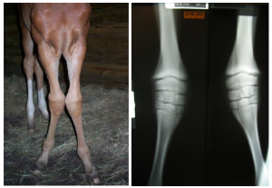
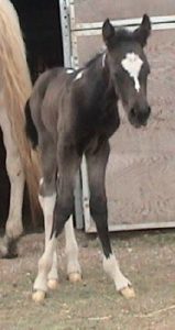
Angular limb deformities are changes in the frontal plane. Angular limb deformities (ALDs) are named according to the joint that is deviated and describe how the distal limb is moving compared to the proximal limb. (Stand at the shoulder or hock and look down to see what joint is deviating).
- Valgus = deviation to the lateral side (note both “L”s)
- Varus = deviation to the medial side
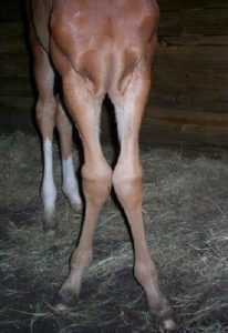
Windswept foals have both a valgus and varus deformity:
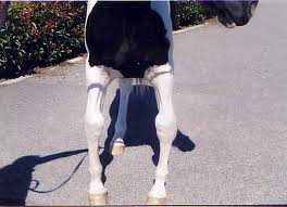
The most common versions are carpal valgus, tarsal valgus and fetlock varus. Mild carpal valgus can be protective; angulation less than 5 degrees in a foal is considered normal. Varus is problematic and needs correction.
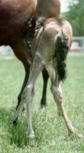
Many foals have external rotation of the forelimbs. The toes point outward. However, the limb is straight but is rotated from the shoulder.
This will correct as the horse develops a broader chest. It is important to not diagnose or misdiagnose an ALD if the limb is rotated but straight. Line up with the foot and/or look down from the shoulder.
Not all ALDs come from a physeal abnormality. Other causes include ligamentous laxity and cuboidal bone abnormalities. Newborn foals can just have weak ligaments. These limbs can be straightened manually and you may be able to see changes as they move. These foals should not be lame.
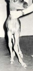
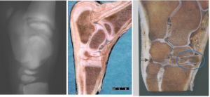
Premature foals often have immature cuboidal bones (the carpal and tarsal bones are still mostly cartilage). Immature bones squash and then ossify in an abnormal shape.
Diagnosis
To determine the cause of the ALD, radiographs are taken using long plates. Draw a line down the middle of each long bone. Where they intersect is typically the cause of the problem (joint or physis). Radiographs also let you assess the cuboidal bone development.
Treatment
Different physes grow at different rates and for different times. Eventually the physis “closes” and further growth is not possible. For correction to work, the physis still needs to be growing. Monitor the more distal joints closely as you have a smaller window of growth.
- Distal radial physis – most growth by 6 months; closes at 3.5 years.
- Distal tibia – most growth by 4 months; closes at 2 years
- Distal MC III/MTIII – most growth by 2 months; closes at 1.5 years
-

https://equineink.com/2019/08/10/the-stages-of-equine-skeletal-development/ .These timelines are more related to radiographic closure; functional closure is much earlier (eg 2 months for the pastern area vs 6 months)
Medical management requires active growth!
- foals with immature cuboidal bones need aggressive support; refer these. They need to be kept recumbent to avoid weight bearing and will often need special casts to keep the limbs straight when they start to move around
- control exercise
- Some weight bearing is fine but more exercise is more trauma. Stall rest is generally indicated.
- adjust the load on the foot through trimming or hoof wall extensions (eg glue on shoes with a wing or extension to one side)
- varus – trim medial aspect or add lateral extension
- valgus – trim lateral aspect or add medial extension
- trim to the side of the deformity or put extension on the opposite side
- increase load on the affected side so increases bone growth
- more effective for fetlock (whether you want it to be or not)
- varus – trim medial aspect or add lateral extension
Surgery is needed if
- severe angulation (>15 degrees)
- not enough growth potential left for correction to occur naturally
- foal is developing a secondary conformational abnormality (foal with carpus valgus is developing a fetlock varus)
- economically important
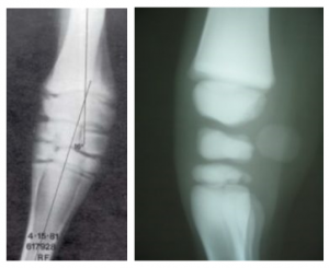
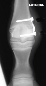
Implants are put on the faster growing side to slow the growth on that side and let the other side “catch up”. It is possible to overcorrect so implants need to be removed as soon as the limb appears straight. Historically we performed periosteal stripping to stimulate the slower growing side. These may or may not have done anything (mother nature fixed them?) but overcorrection was not an issue. Implants may be a combination of screws and wire as seen in this image (transphyseal bridging) or a single screw placed across the physis (transphyseal screw).
Remember: These implants need to be removed if the limb becomes straight and the foal is still growing as the limb can overcorrect.
Key Takeaways
Angular limb deformities can occur due to ligamentous laxity in newborn foals, due to crushed cuboidal bones (due to prematurity and poor ossification), or due to trauma and/or inflammation of the growth plate. Mother nature will fix some of these but success depends on the physis involved and the degree of angulation. If the fetlock is involved, surgery is indicated earlier rather than later as this physis closes very early in life. Newborn foals with ALDs should be radiographed to assess cuboidal bone maturity.
Resources
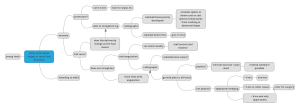
Malone’s flowchart for ALDs (ALD mindmap pdf)
Single-incision drilling technique to achieve hemiepiphysiodesis of the distal metacarpus – complications and outcome in 207 foals with
metacarpophalangeal varus deformities. Veterinary Surgery. 2023;52:26–32. – it worked well
