Large animal cutaneous neoplasia
How to – Mass Removal
Indications
Mass removal is indicated for known and suspected tumors. Excisional biopsy works well in many situations in which full mass removal is possible. Portion of the mass is submitted for histopathology. Follow-up treatment may or may not be needed.
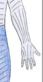
Relevant anatomy
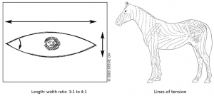
Masses should be removed along the lines of tension.
Preoperative management
Food restrictions: NA if performed standing. If under general anesthesia, food should be withheld for 6-12 hours in horses and 24-48 hours in adult ruminants.
NSAIDs/analgesics: Preoperative NSAIDs or narcotics are used to prevent pain windup.
Antibiotics: Often not required. Preoperative antibiotics may be indicated in contaminated environments or when surgery involves the GI tract, respiratory tract or urogenital tract.
Tetanus prophylaxis is recommended in horses.
Local blocks: Regional blocks are often used to circumscribe the mass to be removed.
Position/preparation: Standing sedation is preferred when possible. Local blocks may be all that is required in cattle. Presuturing for 2-8 hours may help stretch skin for larger masses.
Surgery Supplies:
- Scalpel and handle
- Hemostats
- Metzenbaum scissors
- Thumb forceps
- Needle holders
- Suture – 2-0 to 3-0, cutting needle for skin
- Suture – 2-0 to 3-0 taper needle for subcutaneous tissues
- Penrose drain (optional)
- Formalin for histopathology
Surgical procedure
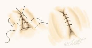
- A fusiform incision is made around the mass, long axis along the lines of tension. The length should be approximately 3-4x the width needed to remove the mass.
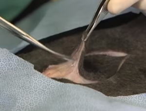
- The subcutaneous tissues are dissected, freeing the mass from the underlying tissues.
- Subcutaneous closure may be skipped; alternately a simple continuous pattern is used to close deadspace.
- If the deadspace is large, a penrose drain may be placed.
- Skin closure
- Routine – simple continuous or Ford interlocking
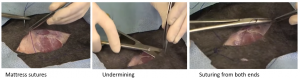
- Options for closure under tension
- Start closing from both ends
- Tension relief sutures in key parts – NFFN, vertical mattress or horizontal mattress
- Undermine tissues to allow stretch
- Relief incisions
- Secure knots by first placing granny knots or using 3 throws rather than 2 for the surgeon’s knot. Follow with a square knot. The granny knot can be tightened with the later throw meaning you can tighten down in two steps. Three throws provides more friction that keeps the knot in place.
- Place a stent over the incision or bandage the area if needed to compress deadspace.
Postoperative care
- Minimize motion for 3-5 days and then limit motion until suture removal.
- Suture removal in 10-14 days.
- NSAIDs for 3 days will help minimize swelling
Complications
- Dehiscence (usually about day 3)
- Infection
- Tumor recurrence
- Poor healing (tumor cells at wound margin)
Videos
Mass removal and closing wounds under tension
standing version
Resources
Using a callicrate bander to remove a pedunculated mass on a bovine – video
sutured edges come apart; lack of healing of incision
