Bovine Lameness and Podiatry
How to – Digit amputation
Indications
Bovine digit amputation is recommended when the digit is painful and unlikely to improve, deep digital structures are infected, or when drainage is needed for the flexor tendon sheath.
The lateral digit on the hindlimb and the medial digit on the forelimb are the main weight bearing structures. Removal of either of these will significantly impact the lifespan of the animal in the herd. Breeding bulls also need full function of all hind digits. Arthrodesis should be considered instead of amputation in these instances.
There are two basic types of digit amputation. This is one version and enables rapid coverage of the exposed surfaces with granulation tissue.
Relevant anatomy
The amputation should be performed as distally as possible to maintain the supporting structures of the limb. Whenever possible, the amputation is performed through P2 or the limb disarticulated through the pastern joint.
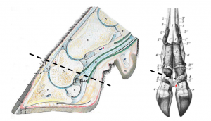
This level of amputation removes any infection in the coffin joint and navicular bursa. It also opens the flexor tendon sheath for drainage.
Preoperative management
Food restrictions:
The procedure is usually short enough that food restriction is not required. If more surgery time is anticipated, food should be withheld for 48 hours whenever possible.
NSAIDs/analgesics: Preoperative and postoperative analgesics are recommended.
Antibiotics: Not usually required.
Local blocks: IVRA (iv regional anesthesia) or ring block
Position/preparation: The cow is cast into lateral recumbency (with the affected digit accessible) using sedation and casting ropes.
Surgery Supplies:
- Gigli wire and handles
- Wire cutters
- Scalpel blade (no handle)
- Mosquito hemostats
- Bandage material
Surgical procedure
- Using the scalpel blade, make a sharp incision to the bone on the axial aspect of the digit. The incision is made above the coronary band. Keep the incision as distal as possible while still ensuring the infected or traumatized tissues will be removed
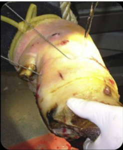
- Seat the gigli wire in the incision and use the handles to saw through the bone
- Saw at an angle so the surface of the bone is angled with the higher side more abaxial. This will minimize the risk of the cow getting her digit stuck someplace
- The scalpel may be needed to finish cutting though the skin on the abaxial aspect.
- If cartilage is exposed, it should be debrided to permit granulation tissue coverage.
- Remove any small fragments of bone.
- Use the mosquito hemostats to clamp, twist and pull (Cornell cautery) any bleeders.
- Bandage the foot, being careful to not add so much bandage that the cow is walking on the bandage.
Postoperative care
- The bandage is typically left in place for 5 days (until granulation tissue can cover the exposed bone).
- Change the bandage if it becomes soaked through with blood or if it becomes wet or soiled.
- Remove the bandage if still in place 10 days postoperatively.
Complications
- Exposed cartilage may prolong healing time (but it is a cow, so not by much)
- Cattle may have difficulty on slatted floors or with prolonged standing.
- Breakdown of the opposite digit is expected. Cow life expectancy in the herd is <18 months.
Videos
Resources
An Update on the Assessment and Management of Pain Associated with Lameness in Cattle VCNA FA 2017; Volume 33, Issue 2, July 2017, Pages 389-411
put into recumbency using ropes
