Bovine Lameness and Podiatry
Approach to the lame cow
The vast majority (85%) of bovine lameness originates in the foot. The lateral hind digit is the most common site.
There are typically three main options for a lame bovine:
- cull
- used if the lameness is severe, the animal has other issues (poor body condition etc), the disorder has a poor prognosis or will be difficult to treat (eg joint infections)
- refer or treat with surgery
- referral is rare as it is costly in both time and money and opens the herd to biosecurity issues
- surgery options will depend on the comfort of the practitioner and the farm environment (management, housing etc)
- treat on farm
- trimming
- analgesics
- supportive care
- +/- antibiotics
This means the first step is to determine if treatment on the farm is a viable alternative. Many foot lesions can be managed on the farm; most conditions above the foot are more difficult to manage.
Foot issues that can require more intensive management or surgery include those with damage to the coffin bone and/or deep seated infections. Digit amputation, arthrotomies and/or arthrodesis can be successful in managing these but do take repeated intensive treatment.
Above the foot, arthritis can be treated with analgesics and supportive care. However, arthritis is rarely an identified concern except in breeding bulls. Most other conditions above the foot are difficult to manage, particularly in adult animals. These include damage to joints, infection in synovial structures, tendon/ligament injuries, nerve injuries and fractures.
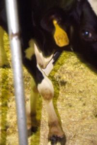
Non-weight bearing lameness
Cattle are not built to maintain weight on three limbs; non-weight bearing lameness needs to be evaluated and treated aggressively or animals euthanized (they cannot be sent to slaughter unless ambulating on all four limbs).
Differentials include
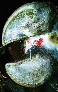
- White line infection
- Fracture
- Joint luxation
- Injury of a weight-bearing ligament or tendon (eg, gastrocnemius muscle)
- Nerve injury affecting weight-bearing (eg, radial nerve, femoral nerve, sciatic nerve)
- Septic arthritis or tenosynovitis
Of these, white line infections have the most favorable prognosis. The remaining conditions are challenging to treat and have a guarded prognosis. While very valuable animals (any condition) and neonates with certain fractures are treated, most cattle with bone/joint/nerve/tendon injuries are euthanized.
Determine if the pain is in the foot or above the foot.
Note: Almost all conditions start as weight-bearing and progress to non-weight bearing. These are conditions that might rapidly move to non-weight-bearing.
Lameness localization
A physical examination is key to most lameness localization in cattle. The foot needs to be assessed closely for injury and lesions.
Hoof testers can be applied. Changes in stance can give clues about what structures are painful (eg standing on toes if heels hurt). The front limbs may be crossed if the cow is attempting to avoid weight bearing on the medial digits.
If the foot is normal, the remainder of the limb is assessed for swelling and/or pain.
Nerve blocks are not routinely performed. Intra-articular blocks can help with evaluating lameness above the foot. If needed, the foot can be numbed with a “ring block” (local anesthetic injected circumferentially around the limb). The ring block is typically performed midcannon bone as this is narrowest part of the limb.
It is also possible to numb the lower limb with an IV regional anesthetic block (IVRA). A tourniquet is placed around the cannon bone and local anesthetic (usually lidocaine) is placed into a vein below the tourniquet. The lidocaine diffuses into the tissues and blocks sensation. The effect wears off as soon as the tourniquet is removed. Unfortunately, the tourniquet itself can cause pain so the interpretation can be tricky. IVRA is used primarily for treatments rather than diagnostics.
Radiographs and/or ultrasound are useful to further explore injuries and swellings.
Key Takeaways
85% of the time, the lameness is in the hoof. Over 70% of those are in the lateral digit of the hindlimb, particularly in dairy cattle.
A midcannon ring block is typically used to numb the foot if no lesions are identified on gross examination.
If the lesion is above the foot, the prognosis is generally guarded.
Foot lesions can be challenging to treat but many respond to trimming and management changes.
Resources
The Impact of Lameness on Welfare of the Dairy Cow, VCNA FA 2017; Volume 33, Issue 2, July 2017, Pages 153-164
An Update on the Assessment and Management of Pain Associated with Lameness in Cattle VCNA FA 2017; Volume 33, Issue 2, July 2017, Pages 389-411
The Dairyland Initiative Lifestep Lameness Module
Diagnosis and therapy of feedlot lameness, AABP 2020
inflammation of a tendon sheath
