Bovine Lameness and Podiatry
Foot sepsis
What is it?
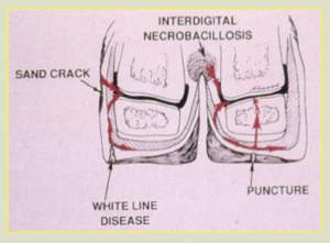
Several structures in the digit can become infected, typically due to ascending infections from foot rot or foot abscesses that develop from sole ulcers or white line disease. Interdigital trauma can also be a source of infection. The pedal bone (P3), the navicular bone, the coffin joint and the flexor tendon can be impacted, often in various combinations.
How to recognize it?
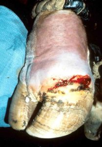
Affected cattle are usually non-weight bearing lame and the lower limb and coronary band are often swollen asymmetrically.
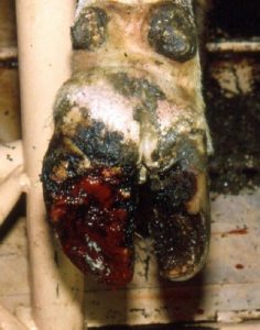
Fistulous draining tracts may be evident above the digit.
The foot is often sensitive on hoof tester examination and other pathology typically visible on foot examination. The lameness improves with IVRA (regional anesthesia).
Pathogenesis
Bacteria involved are typically those in the environment and related to fecal and skin flora. Mixed populations are common and include E coli, Trueperella pyogenes, Staphylococcus and Fusobacterium. Infection ascends due to the hard sole and limited drainage.
Bacterial infections elsewhere in the foot lead to infection of deeper structures
Diagnostics
Confirmation of joint sepsis is obtained by passing a probe into the joint or synovial structure through a fistulous tract or through sampling of joint fluid (infected if >40K cells/ul). Cow pus is thick; a 14ga needle may be necessary to obtain a sample. Radiographs are useful to identify bony changes. Bone lysis can be seen with bone infection and periosteal reaction with infection near the surface of the bone. Changes develop faster in cattle than in horses but still take 4-14 days to be visible radiographically.
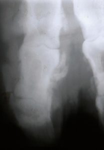
Cow pus is also so thick that it will physically push the bones apart, widening the joint space. Gas may also be present radiographically
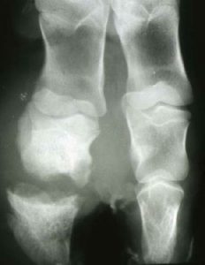
Pedal osteitis is infection of P3 and develops due to similar ascending infection. The local inflammation lead to significant lysis of P3.
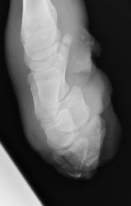
The navicular bone can be similarly affected.
Treatment
Joint lavage via needles or arthrotomy (preferred) is used for acute cases of synovial infection. Cattle rapidly develop fibrin within the joint, making needle lavage of limited use. Systemic antibiotics are indicated.
With chronic infection in the distal limb (pastern or coffin joint), lavage and antibiotics are minimally effective. Treatment options are amputation of the digit or arthrodesis the joint (facilitated ankylosis). Amputation works best in unilateral lesions in smaller stature animals that are not housed on slatted floors. Amputation is the fastest means of removing pain and infection. Survival is limited. On average, the animal will last ~ 18 months in the herd before they are culled for related issues. Arthrodesis is generally needed for bulls, heavy animals, uneven flooring, and valuable animals. Referral or consultation is recommended if arthrodesis is desired.
Bone infections are treated by debridement and/or digit amputation.
Key Takeaways
A non-weightbearing bovine may have deep digital infection – in the joint, bone or tendon sheath.
- Antibiotics and/or joint lavage are rarely successful in treating these conditions by themselves
- Digit amputation is a means of rapid removal of the problem but does limit lifespan of the animal due to breakdown of the opposite digit
- Ankylosis involves debridement and fusion of the infected joint and can lead to a full lifespan in larger or valuable animals; it does take more time to resolve the pain.
Resources
Lameness originating in the hoof of cattle, Cramer and Solano, merckvetmanual.com
Surgical Procedures of the Distal Limb for Treatment of Sepsis in Cattle; VCNA FA 2017; Volume 33, Issue 2, July 2017, Pages 329-350
An Update on the Assessment and Management of Pain Associated with Lameness in Cattle VCNA FA 2017; Volume 33, Issue 2, July 2017, Pages 389-411
