FA GI Diagnostics & GI Surgery Principles
Abdominal ultrasound
Ultrasound is a valuable tool for identifying peritoneal and GI tract abnormalities in cattle. Many structures look similar when normal; location is an important key as is knowing the normal sonographic appearance. Most of these evaluations can be performed with a linear probe.
Start by scanning ventrally for fluid pockets. Check to the right of midline and under the flank fold, just beside the udder.
Left side evaluation
Left side structures are the reticulum, rumen, and spleen.
Reticulum
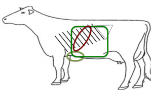
The reticulum can be visualized both the left and right of the sternum, ventral to the point of the elbow.
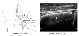
The reticulum normally has a half-moon shape. Due to the gas content, foreign bodies and magnets are not visible. In this diagram, 1 is the reticulum, 2 is the craniodorsal blind sac of the rumen, and 3 is the ventral sac of the rumen. Regular contractions that can be evaluated to determine whether motility is normal. The reticulum should have one biphasic contraction per minute; this is typically immediately followed by a rumen contraction.
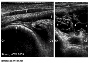
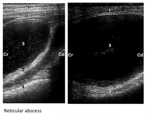
Scan the caudoventral reticular wall to evaluate for hardware related reticuloperitonitis and reticular abscesses.
Rumen
The rumen is visible along most of the left side. The gas cap means only the external layer is visible dorsally. More fluid contents can be seen ventrally.
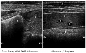
Spleen
The spleen has a hyperechoic texture with prominent vessels. Occasional abscesses can be observed within the spleen.
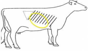
Right side evaluation
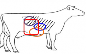
Structures on the right side include the omasum, abomasum, small intestine and large intestine.
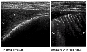
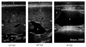
Omasum
The omasum is a crescent shaped structure on the right, in the 6th to 11th intercostal spaces (ICS). It is easiest to see in the 9th ICS since it is closest the body wall in the 8th-9th ICS. The omasum should not contract. The leaves are not visible unless there is fluid reflux from the abomasum.
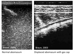
Abomasum
Small Intestine
Small intestine is visible ultrasonographically from the tuber coxae to the 8th ICS ranging from the transverse processes dorsally to the midline ventrally.
The duodenum is wrapped in omentum in the central aspect of the right paralumbar fossa.
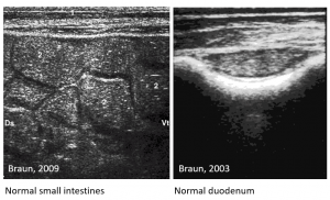
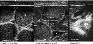
Detectable abnormalities include intestinal ileus or motility disturbances, hemorrhagic bowel syndrome and intussusceptions.
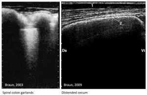
Large Intestine and Cecum
The large intestine is a gas filled structure medial to the descending duodenum. The spiral colon will often have a garland appearance. The cecum is nondescript but can be identified when distended and resonant.
Liver
The liver is visible ultrasongraphically on the right, extending from just caudal to the last rib up to the 5th ICS. It is often partially obscured by the lung.
Normal liver is fairly homogenous with visible gall bladder and biliary tree.
GI Challenge
Play concentration with lesions and ultrasound images – Level B
Resources
Stieger-Vanegas, McKenzie. Abdominal imaging in small ruminants. VCNA 37 (2021) 55-74. Worth saving for later use!
Braun. Ultrasonography of the Gastrointestinal Tract in Cattle. VCNA 25 (2009) 567–590
Braun. Ultrasonography of the Liver in Cattle, VCNA 25(2009) 591-609
Braun. Ultrasonography in gastrointestinal disease in cattle. The Veterinary Journal, 2003, Vol.166(2), pp.112-124
