FA GI Topics
How to – Right paramedian abomasopexy
Indications
Stabilization of the abomasum is part of any DA surgery. Right paramedian abomasopexy is one option. This is a direct tack of the abomasum in its normal location. This method can be performed in cows that cannot stand due to musculoskeletal trauma or milk fever.
Relevant anatomy
The abomasum is normally located on the ventral abdomen, typically just to the right of midline. It often shifts horizontally across the midline during late gestation. When gas filled, the abomasum can displace to either the left or the right sides of the abdomen and float up. The pylorus will stay more ventral while the body of the abomasum floats more freely upward.
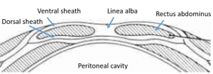
The ventral abdominal wall is comprised of the continuation of the flank muscles; however these turn into fascia. Midline is the linea alba. Off midline is the rectus abdominus muscle encased between the dorsal and ventral rectus sheaths. The ventral rectus sheath is the strength layer.
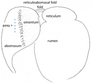
Exploratory options are limited from this approach due to the massive rumen. The abomasum should sit between the reticulum and rumen.
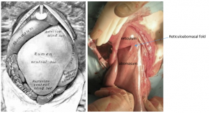
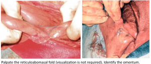
The abomasum is identifiable by the noticeable mucosal slip as the wall is palpated. The reticulum has a honeycomb texture and is connected to the abomasum by a ligament (reticuloabomasal fold).
Omentum attaches to the greater and lesser curvatures of the abomasum. In this area, the omentum contains minimal fat and is best identified by the vascular arborization evident where it attaches to the abomasum.
Preoperative management
Food restrictions: Cows with a displaced abomasum are generally off feed already (poor appetite).
NSAIDs/analgesics: Recommended preoperatively. Flunixin meglumine 1.1-2.2 mg/kg iv is standard.
Antibiotics: Recommended. As cows can wall off infection, there can be surprises inside. If the cow is milking, ceftiofur 2.2 mg/kg im is standard. In beef or non-lactating cattle, other options do exist. Remember: the label dose of procaine penicillin is ineffective!
Local blocks: U block (line block in a U shape) or line block
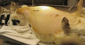
Position/preparation: The animal is positioned in dorsal recumbency. This typically involves sedation (20 mg xylazine or similar) and casting ropes. The gutter makes a decent trough to help hold the patient in dorsal; bales can also help to support her in that position.
Surgery Supplies – Additional
- 2 PDS or nylon for the pexy and the ventral sheath
- 0 chromic gut or similar for the muscle closure
- 3 vetafil or similar for skin closure
Surgery Supplies- Standard
- Standard surgery pack
- Sterile sleeves for internal palpation
- Scalpel blade and handle
Surgical procedure
- In an adult cow, a 6-8″ incision is made approximately 4″ to the right of midline and approximately 4″ caudal to the xiphoid. This is the normal location of the abomasum. In smaller animals, adjust the distances for relative body size. If the milk vein is in the intended site, make the incision closer to midline.
-
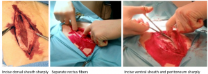
- The incision is made through skin, external sheath, rectus abdominus, internal sheath and peritoneum
- Blunt dissection should be performed through the rectus abdominus (less bleeding)
-
- Any vessels encountered should be ligated; bleeding is typically minimal during surgery but that can change dramatically when the cow returns to her feet.
- If indicated, exploration can be performed. Sterile sleeves help minimize contamination. Exploration is needed if the abomasum is not immediately visible. The abomasum usually floats to its normal position. If it does not, adhesions are likely – either from abomasal rupture, peritonitis or previous abomasal tack.
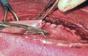
- If the abomasum is in its normal position, the pexy can be performed. The objective is to incorporate the wall of the abomasum in the closure of the dorsal rectus sheath.
- Identify the cranial aspect of the abomasum by locating the reticuloabomasal fold. This ensures the pexy is not near the pylorus.
- Identify the attachment of the greater omentum. The omentum should be avoided in the pexy. The pexy should directly incorporate the greater curvature of the abomasum.
-
- The pylorus does not need to be exteriorized or visualized in most cases.
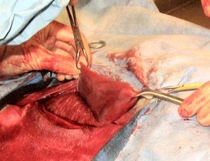
- Exteriorize 6-8″ of abomasum using wet towels to grasp and lift it out of the incision ~2″. Identify the pexy site. This should start about 3″ caudal to the reticuloabomasal fold and abaxial (lateral) to the omental attachment.
-
- Slip the mucosa away and place a towel clamp at either end of the exposed abomasum. This will allow you to move the abomasum in and out of the abdomen as needed.
-
- Start the pexy by placing a suture (#2 PDS or nylon) across the caudal aspect of the dorsal sheath. Secure the bite with a knot. There is no need to include the abomasum in this bite.
-
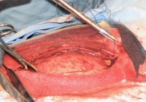
- Close the dorsal sheath using a simple continuous pattern. Incorporate a bite of abomasum in each pass, making sure the mucosa is not included in the closure (slipped away).
-
- After 6-8 bites, close only the ventral sheath. Stop including the abomasum.
- Close the muscle using 0 gut, simple continuous pattern.
- Close the external sheath using 2 PDS or nylon, simple continuous pattern. Do NOT include muscle. When it tears loose, your closure is now loose too.
- Close the skin using 3 Vetafil, Ford Interlocking pattern.
- Return the cow to sternal recumbency/standing.
Postoperative care
- Suture removal in 10-14 days.
- Monitor the cow for any signs of infection or peritonitis.
- Consider rumen transfaunation if available.
Complications
- Poor oxygenation (maximum surgery time ~ 1 hour)- Pa02 drops from 86% standing to 65% at 15 min and 61% at 30 min
- Incisional dehiscence (rare)
- Incisional infection (uncommon) or hernia (rare)
- Subcutaneous hemorrhage (uncommon) – apply a tight belly band for 24-48 hours
- Peritonitis
- Abomasal fistula (if suture went into lumen)
- Recurrence (generally due to suture breakage)
Videos
sheep abomasopexy annotated- video
