Equine Oral, Esophageal and Rectal disorders
Dental surgery
Equine teeth are firmly packed together and develop issues readily. Miniature horses have regular sized teeth. Horses develop habits of chewing on things. In other countries, horses are fed silage and don’t brush afterwards. Diseases lead to extra calcification and pain or to tooth loss.
Dental disorders (tooth root infections) are also one of the most common reasons camelids are evaluated by veterinarians.
The biggest thing we worry about is whether or not to remove a tooth. It may be diseased due apical infection, fractured (removal may not always be required) or in an abnormal position (malalignment). More and more, we are removing incisors to treat ETORH (equine odontoclastic tooth resorption and hypercementosis).
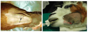
Clinical signs associated with dental disease include nasal discharge or fistula (maxillary teeth) or draining tracts (mandibular teeth). Horses may also have malodorous breath and quidding (abnormal chewing).
CT scans are ideal for diagnosing dental disorders. Radiographs are often confusing!
Tooth removal methods
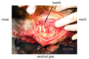
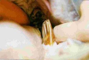
Oral extraction
This is the best option if possible. Oral extraction carries the fewest complications. However, it takes patience and skill due to the tightly packed teeth. If the tooth enters the sinus (most do), it needs to be combined with sinus flush.
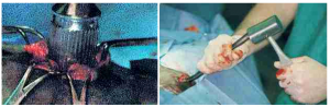
Tooth repulsion
We used to remove teeth through repulsion. with the horse under general anesthesia, the tooth was “hammered” out from the root surface. The hammering loosened the tooth so it could be pushed or pulled out of the socket. Root access was gained by trephination or sinusotomy. This method was very traumatic to adjacent structures; complication rates were close to 50%. Tooth repulsion has not been used in camelids due to the narrower and more easily fractured mandible.
Intraoral tooth removal + repulsion
Buccotomy
Buccotomy is an alternative option for removal of cheek teeth. The tooth is approached through the side of the jaw. This method is commonly used in camelids due to the thinner, more easily fractured jaw. This approach requires specialized drill bits.
Postoperative care
After tooth removal, the socket may be left open to fill in with granulation tissue. With this method, the socket should be irrigated daily to remove food material until healed. This can take awhile. Care is long and tedious; sometimes it doesn’t heal and horses are left with a fistulous tract. Alternately, the socket can be filled with a temporary seal for 4-6 weeks in order to prevent food contamination of the area. Sealants include gauze packing, wax, polymethylmethacrylate (bone cement) and gutta percha. The seal should be seated so it occupies only the proximal 20% of socket. The rest fills in with granulation tissue. As the granulation tissue develops, it is supposed to push the sealant out of the socket. Occasionally manual removal is required. Finally, a permanent prosthesis can be used. Often polymethylmethacrylate is used as a permanent tooth replacement. This helps prevent overgrowth of the opposite tooth and subsequent mastication problems. However, some will create a chronic fistula as a foreign body. Without a placeholder, the adjacent teeth will drift into the socket and the opposing teeth will overgrow.
If the sinus was involved, sinus lavage is also necessary.
Complications
Complications with tooth removal include:
- damage to adjacent teeth
- damage to the alveolar bone
- removal of the wrong tooth
- retained root fragments
- dry socket
- fistula formation
- osteomyelitis
- continued nasal discharge
Expected ongoing care is related to the overgrowth of opposing teeth and the shifting of adjacent teeth. Horses with a removed tooth require frequent dental equilibration.
Alternative procedures
If surgical removal isn’t indicated or isn’t an option, management of the the sinusitis or infection may help. The sinus can be flushed through trephination and insertion of a catheter lavage system. Flushing through a 28-32Fr catheter once or twice daily can help clear the infection and may be less costly in some cases.
Endodontic treatment is possible but not commonly performed. “Root canals” can only be performed if the periodontal ligament is intact. With a root canal, the diseased pulp is removed and the cavity sealed. Periapical curettage may be sufficient with mandibular tooth infections. It is not recommended if the tooth is cracked below the gum line, an oral fistula is present, or the horse has a deep corner periodontal pocket.
Resources
Complications of equine cheek teeth extractions Eq Vet Educ 2023, 35:384-392
Dental Disorders of Donkeys, VCNA 2019, Vol.35(3), pp.529-544
Equine Odontoclastic Tooth Resorption and Hypercementosis, EVE 2018, Vol.30(7), pp.386-391
