Bovine musculoskeletal disorders
Synovial structure disorders
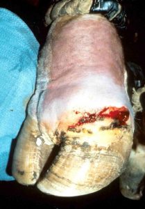
Septic synovitis (joints, tendon sheaths and bursas)
Joint infections are a common cause of lameness in neonates and young feedlot animals.
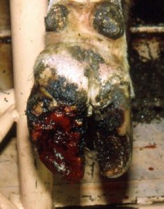
Infections can enter via direct inoculation (wound or injection), local extension of infection (eg via hoof ulcer or foot rot) or via the blood supply (infection elsewhere). Hematogenous spread is most common in neonates, especially for the larger joints, but can be seen in all ages and can affect multiple joints. In calves, synovial infections are typically associated with umbilical infections, respiratory infections or GI infections. Adults can get hematogenous spread, especially to sites of injury, even if there is no wound.
Animals are typically non-weight bearing and the area is often swollen and painful.
Cytology can be useful but cows produce lots of neutrophils. Consider a synovial structure infected if >40K cells/ul. Colostral antibodies should be evaluated in neonates.
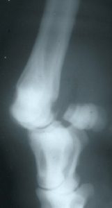
Bony changes develop fast in cattle but still take 4-5 days to appear on radiographs. If there is infection near bone, it will develop extensive periosteal reaction (except P3, no periosteum). This can be a useful clue.
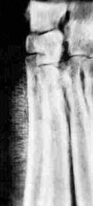
Bones will also lyse when infected. Cow pus is so thick that it will physically push the bones apart, widening the joint space.
Treatment is high volume lavage (generally need arthrotomy), NSAIDs and four weeks of systemic antibiotics. Animals will generally need restricted exercise for pain control. Physical therapy may be required to minimize limb contracture, especially in young animals. [Limb contracture occurs due to a pain response, similar to touching a hot stove. The body will withdraw (contract) the limb reflexively. Particularly with young animals, it can “freeze” this way.]
*In cattle, you just end up flushing the tract with needle lavage. The fibrin is so thick that you can’t flush it out through a needle. Arthrotomies are often necessary to actually flush the junk out – at least in my hands. EDM.
With chronic infection in the distal limb (pastern or coffin joint), you can amputate the distal limb or arthrodese the joint. Amputation works best in unilateral lesions in smaller stature animals that are not housed on slatted floors. Survival is limited. On average, the animal will last ~ 18 months in the herd before they are culled for related issues. Arthrodesis is generally needed for bulls, heavy animals, uneven flooring, and valuable animals. Referral is recommended if arthrodesis is desired.
Prognosis depends on rapidity and effectiveness of early treatment. In calves, the cause of the infection also needs to be addressed (umbilical resection, etc).
Osteochondrosis
Osteochondrosis is much less common in cattle. [See equine osteochondrosis chapter as a resource.] It is seen in bulls and fattening steers. Generally it is not treated.
It is very common in swine and is not treated.
Hygromas
Hygromas are extra-articular swellings that occur on the lateral aspects of the hocks or the dorsum of the carpus. These may reflect poor bedding or housing issues and occur due to repeated trauma. They are not an issue unless they become infected. Removal is difficult. Drain abscesses.


Cruciate injury
tightening of the tendons leading to a bowing effect of the limb
defect in cartilage maturation process
