LA Respiratory Issues
Pleural space disorders
In cattle, pleural abscesses and pericarditis occur secondary to hardware disease. Prognosis is grave and treatment is not recommended. The surgery is cool, though, and is aided by the bovine complete mediastinum which generally prevents bilateral pneumothorax.
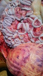
Pleural abscesses in horses are generally secondary to pleuritis and can be treated through drainage once consolidated. Generally pleural surgery is best performed in a referral hospital due to the risk of bilateral pneumothorax and collapse.
Thoracic wounds
Thoracic wounds are the most common cause of pleural space disease in horses. A wound between the ribs can enter the thoracic cavity, the peritoneal cavity or both. Due to the curve of the diaphragm, any wound after the 6th intercostal space could enter the peritoneal cavity; this often means both the thoracic and peritoneal cavities should be evaluated carefully using ultrasound, radiographs, and/or fluid evaluation. With trauma, pneumothorax, pleuritis and peritonitis can develop with significant consequences. Horses with pneumothorax show signs of distress with rapid and often shallow breathing. Pneumothorax requires air removal and prevention of more air into the cavity. Trying to remove the air in the field is challenging! A wound may be sutured temporarily or bandaged with an impermeable product until the horse reaches the hospital. A stent bandage (bandage sutured in place) can be easier to place than banding a horses full thorax.
Pleuritis can be very painful. It is treated with antibiotics and/or abscess drainage. Both peritonitis and pleuritis can be fatal.
Axillary wounds are also common and can lead to pneumomedistinum and subsequently to pneumothorax:
Axillary wounds in horses and the development of subcutaneous emphysema, pneumomediastinum and pneumothorax
Summary
Equine axillary wounds are common in horses. Severe and potentially life‐threatening complications that can result from axillary wounds include subcutaneous emphysema, pneumomediastinum and pneumothorax. This report describes the occurrence of these complications and appropriate treatment. Case records of 7 horses after sustaining an axillary wound are reviewed. Of these cases, all 7 developed subcutaneous emphysema, 5 developed a pneumomediastinum and 4 developed a pneumothorax. The time between the wound occurrence and the development of subcutaneous emphysema was able to be determined in 5 of the 7 cases. The mean ± s.d. time for the development of subcutaneous emphysema following initial injury was 3.2 ± 0.84 days (range 2–4 days). Resolution of subcutaneous emphysema was not achieved until the treatment included packing the wound to stop it from acting as a one‐way valve. Horses with a pneumothorax in respiratory distress were managed with thoracocentesis or placement of thoracic drains. Horses with a pneumothorax but without respiratory distress were treated with conservative management. All horses survived to discharge.
Pneumomediastinum has no associated clinical signs.
Neck wounds can also lead to pneumomediastinum. Typically both axillary and neck wounds lead to subcutaneous emphysema which can track into the mediastinum. Typically subcutaneous emphysema is left to resolve on its own; more attention is paid to minimizing its development by minimizing movement of the horse. The horse’s body temperature should also be monitored as subcutaneous emphysema has an insulating effect.
Rib Fractures
Rib fractures can occur in foals during the parturition process. These are often undiagnosed. The fracture ends can cause lung lacerations, cardiac lacerations and diaphragm damage. Approximately 10% of neonatal deaths are due to rib fractures. Diagnosis is made by putting the foal on its back and checking for symmetry and/or by ultrasounding the ribs. Treatment is stabilization. Stabilization is performed by keeping the affected side down, splinting the ribs and with oxygen supplementation. Bandaging is contraindicated with a flail chest (flail =both sides fractured)!
Practice
Level B
Resources
Axillary wounds in horses, EVE Vol 25 (3): 139-143, 2013
How to manage penetrating wounds in the field, AAEP 2012
Thoracic trauma in horses, Volume 31, Issue 1, April 2015, Pages 199-219- denser and more thorough
Treating thoracic injuries, Vet Folio 2009 -worth subscribing
Hardware disease in the bovine, 2015 Acad J of Animal Diseases (we don’t treat as many as they do)
Diagnosis and treatment of hardware disease, 2017 VCNA
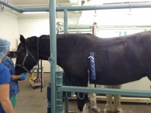
STUDY BREAK
Study break: This is what goes on at our house
