Equine Lameness
Palpation anatomy- hindlimb
Vic Cox
For this exercise we will start distally and then move up the leg.
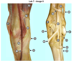
Long and lateral digital extensor tendons
Begin by grasping the upper cannon region and run your thumb over the dorsal aspect of the cannon just distal to the tarsus. You should be able to feel and roll two tendons. The larger tendon is the long digital extensor tendon and the smaller more lateral one is the lateral digital extensor tendon. Trace the two of them distally and note that they fuse in the midcannon region. The lateral tendon is sometimes transected (cut) in order to treat the gait defect known as stringhalt. It is important to note that only the lateral tendon is cut, never the long digital extensor tendon. For that reason, it is important to palpate both tendons to ensure that only the lateral one is cut.
Move up the leg on the lateral side and find the lateral digital extensor tendon above the tarsus where it will be wider than it is distal to the tarsus. As you palpate the lateral digital extensor tendon above and below the hock simultaneously you may be able to feel vibrations in the distal tendon due to palpation of the tendon above the hock.
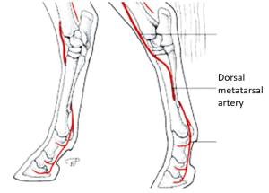
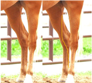
Dorsal metatarsal artery
More laterally, in the upper cannon region, find the groove between the cannon and splint bones. The dorsal metatarsal artery will lie in this groove and must be carefully avoided when cutting the lateral digital extensor tendon. The artery runs under the tendon as it passes distally over the dorsal aspect of the tarsus onto the cannon bone. Careful palpation of this artery in the bony groove will enable detection of the pulse. This artery is an important source of arterial blood for blood gas sampling during surgery.
Medial saphenous vein
On the dorsal side of the tarsus (hock) note the large cranial branch of the medial saphenous vein and palpate it with a light touch. One must be careful to avoid damage to this vein when performing a cunean tendinectomy or tarsal arthroscopy.
Medial malleolus
Medial to the vein note the prominent tuberosity which is the medial malleolus. This important landmark is at the distal end of the tibia.
Medial collateral ligament
Distal to the medial malleolus follow the medial collateral ligament of the tarsus which attaches to the medial malleolus proximally. Dorsal to the collateral ligament find a depression that represents the medial part of the tibiotarsal joint. It is here that the joint capsule pouches out when distended with fluid that is referred to as bog spavin.
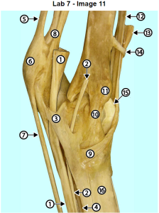
Cunean tendon
Slide you thumb distally over the medial side of the tibiotarsal joint until you feel an oblique tendon which is the cunean tendon. It will lie between the collateral ligament and the saphenous vein and distal to the depression that corresponds to the tibiotarsal joint. The cunean tendon is sometimes cut to lessen the pain of bone spavin (distal hock joint arthritis).
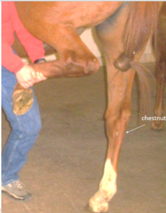
Chestnut
On the distal medial side of the tarsus observe the chestnut and compare its position to the forelimb chestnut.
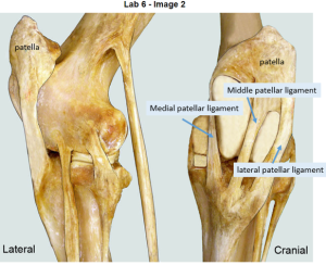
Patellar ligaments
To palpate the stifle joint begin by locating the tibial tuberosity. The tuberosity will be hard and above it the patellar ligaments can be palpated. The middle and medial patellar ligaments are easier to locate than the lateral patellar ligament. This is probably due to the fact that the medial and middle ligaments are part of the loop that forms the patellar locking mechanism. Note the acute angle like the letter “V” formed by the medial and middle patellar tendons where they attach to the tibial tuberosity. Note the tension on the medial and middle tendons and then move your thumb laterally to find the softer, less distinct lateral patellar tendon.
Follow the patellar tendon proximally to the patella. If it is locked, you should be able to feel the large medial part of the femoral trochlea which will be covered with a thin layer of fat. In some cases you will be able to feel the base and then the apex of the patella. Hold the flexor surface of your fingers over the patella and move stifle joint in and out of the locked position. As you do so you will be able the feel the patella locking and “unlocking”.
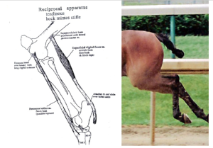
Reciprocal apparatus
Grasp the distal crus on its dorsal aspect and lift the leg in order to flex the stifle joint. As you do so note that the tarsus flexes as the leg is hiked up and the stifle joint flexes. This parallel flexing of the tarsus when the stifle is flexed is evidence of the reciprocal apparatus. With the tarsus elevated pull on the distal part of the limb and note resistance to tarsal extension due to an intact peroneus (fibularis) tertius. This is the test for rupture of the peroneus tertius.
Note that as the tarsus is hiked up and flexes, the fetlock becomes more flexed also. This is due to the action of the superficial digital flexor tendon which is stretched as it passes over the point of the hock (tuber calcis). In contrast, when the hind limb is carried forward in extension, the fetlock is in the extended position as is the hock and stifle.
Resources
Anatomy and radiography of the equine tarsus ibook
Horse anatomy A coloring atlas, Scribd
