Eye surgery
Ocular blocks
Ocular blocks are often necessary for evaluation and treatment. The most commonly performed blocks are the palpebral, supraorbital, and optic nerve blocks.

Horses have such strong eyelid muscles that the palpebral block is required to even examine the eye.
Palpebral block – 1 ml lidocaine, 25ga needle
Lidocaine is injected subcutaneously at the crest of the rostral zygomatic arch or directly adjacent to the nerve as it passes over the arch if the nerve is palpable.
The nerve is a branch of the facial nerve. This procedure blocks motor function – the eyelids can be manipulated. It does NOT block sensation.
Note: we often call this an auriculopalpebral block. Blocking the nerve at either site seems to work.
The supraorbital nerve can be blocked to provide anesthesia of the surgery area during standing procedures or to minimize the need for deeper planes of anesthesia during general anesthesia.
Supraorbital nerve block – 1 ml lidocaine, 25ga needle
The supraorbital foramen can be palpated in the supraorbital rim by placing your thumb and middle fingers on either side of the supraorbital rim. Use the right hand for the left eye and vice versa. Slide your fingers along the rim until you reach the widest part. Drop your index finger down; the foramen is usually directly underneath your finger and is palpable as depression in the bone.
Inject lidocaine over the depression. There is no need to go into the foramen. Injecting the nerve itself is painful and not necessary.
The nerve is a branch of the trigeminal nerve. It provides analgesia to the upper lids. Lower lid anesthesia is most readily performed using a line block.
Optic nerve blocks are required for any enucleation (even those performed under general anesthesia). This is due to the oculocardiac reflex. Traction (pulling) on the optic nerve can lead to life-threatening bradycardia or cardiac asystole due to this reflex. Options include a 4 point block, a Peterson block, and several versions of retrobulbar blocks.
Curved needle retrobulbar block – epidural or spinal needle (20 or 18ga) and 10-20cc lidocaine
Wearing sterile gloves, bend the needle (stylet in place) into a C shape to match the shape of the bony orbit. Insert the needle through the skin between the globe and the bony orbit. As it is advanced, the needle follows the curve of the orbit to the back of the eye and ocular nerve. Lidocaine is injected at the back of the eye until the eye feels turgid and as the needle is withdrawn.

For a 4 point block, 1″ needles are inserted between the orbit and the globe at 4 points evenly distributed around the eye. 5 cc of lidocaine is injected at each site. This block minimizes trauma but is also less effective.
For the Peterson block, a spinal needle is directed from the notch between the zygomatic arch and supraorbital bone toward the back of the eye. Direct the needle straight in until the mandibular ramus is touched, then redirect the needle toward the back of the globe. Often you can see the eye move in response.
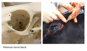
The retrobulbar and Peterson blocks are recommended only for when the eye is being removed as there is a risk of trauma to the optic nerve or artery.
Key Takeaways
Use local blocks for all enucleations.
- Block the optic nerve to prevent the heart from stopping when the eye is being removed
- For standing procedures, block the motor function (muscles) as well as sensation
Resources
4 point block (note this would be easier if they had better syringe handling skills)
Head blocks video: spelling issues but good content
How to prepare for ocular surgery in the standing horse, AAEP, 2002- has pictures
Field surgery of the eye and periorbital tissues, VCNA, 2008 – good for blocks and sedation
These videos also show nerve blocks.
