Large animal wounds
Heel bulb lacerations
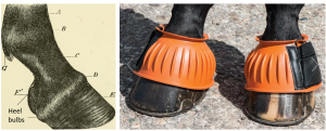
Horses injure their heel bulbs on a regular basis, often by stepping on their own foot but also by clipping the bulbs on nearby structures or when stepped on by other horses. Some horses wear “bell boots” to avoid clipping themselves due to the stride length of the hindlimbs. This is called over-reaching or grabbing.
Heel bulbs are also commonly lacerated when horses kick through structures such as barn walls or sheet metal barriers.
While many mostly involve skin, some can be very serious.
Structural damage
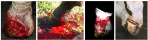
These lacerations may involve the palmar/plantar digital artery, vein and/or nerve and often more than one structure due to the colocalized anatomy (neurovascular bundle). Lacerations of the artery can lead to significant blood loss. If both digital arteries are severed, the foot may become avascular. Most horses will injure one side and essentially give themselves a unilateral neurectomy combined with blood letting.
Synovial structures are abundant in the area and include the digital flexor tendon sheath, the pastern joint, coffin joint and navicular bursa. More than one structure can be lacerated, as well.
Tendon and ligament damage is also possible as is damage to the collateral cartilages. None of these structure heals well due to limited vascular supply.
Finally, the coronary band can be injured, leading to abnormalities in hoof growth.
Therapy
As with other wounds, first ensure the horse is stabilized. Arterial damage needs to be controlled by tight wraps, ligatures and/or tourniquets. More severe wounds should be referred to a hospital at that stage and without worry about wound investigation.
Once the bleeding is controlled and the horse is in a clean environment, regular wound evaluation proceeds with attention to nearby synovial structures, tendons and ligaments. Systemic antibiotics and regional limb perfusion may be indicated to manage the infection risk. Ventral drainage is often challenging due to the firm wall and sole.
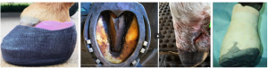
Finally, the heel bulb region is designed to move with each step. Hoof stabilization is often needed to allow wound healing with minimal granulation tissue. With movement, more tissue develops in the wound site, leading to deformities in the horn tissue. The hoof can be stabilized with a bar shoe or with a foot or phalangeal cast.
Prognosis
Prognosis is generally good for return to performance if synovial structures are not involved.
Resources
Equine heel bulb lacerations: 62 cases (2004–2018). J Am Vet Med Assoc. 2022 Jul 20; 260(12):1541-1546.
A review of regional limb perfusion for distal limb infections in the horse. 2021 Equine vet. Educ. 33 (5) 263-277
Traumatic foot injuries in horses: surgical management. 2013 Comp Cont Educ 35(1) E1-E9
