Equine Oral, Esophageal and Rectal disorders
Rectal prolapses
Rectal prolapses are (usually) visible extrusions of rectal or intestinal tissue past the anal sphincter. The length of the prolapsed tissue directly correlates with prognosis and treatment options.
Etiology
Rectal prolapses typically occur due to straining. In horses, this can be straining due to diarrhea, dystocia, intestinal parasites, colic, eosinophilic proctitis, rectal tumor, rectal foreign bodies or due to unknown causes.
Classification
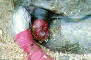
- type I : only rectal mucosa projects through anus
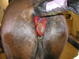
- type II : complete prolapse of all or part of rectal ampulla
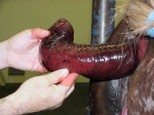
- type III : variable part of small colon intussuscepts into rectum + type II (theoretical only – can’t see)
- type IV : small colon prolapses through anus
Therapy
All involve the 3 R’s – reduce, replace, retain. In other words, reduce the swelling, replace the prolapsed tissue into the rectum, and keep the tissue in place.
Type I prolapses with minimal swelling
Push it back into the rectum. Treat the primary problem. Some of these will return to the rectum on their own when the animal stands up or stops straining.
Type I or type II prolapses with prolonged exposure but no devitalized tissue
- Reduce edema with topical glycerin, sugar, and/or magnesium sulfate.
- Administer an epidural to decrease straining and allow correction.
- Prevent recurrence with loose purse-string suture using umbilical tape. If the purse-string suture is tolerated, leave it in place for 24-48 hours. The purse-string suture should be opened every 2-4 hours to allow rectal emptying.
- Do not feed for 12-24 hours
- Give mineral oil intragastrically or via enema
- Administer laxative diet for 10 days
Type I or Type II with devitalized tissue
- Follow steps for non-damaged tissue
- Perform submucosal resection if partial thickness damage or rectal prolapse amputation if full thickness damage
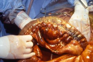
Type III/IV
- monitor abdominal fluid for evidence of ischemic damage.
- laparoscopy or endoscopy of bowel lumen or colostomy
Unfortunately, these usually strip the blood supply off the small colon when they occur.
Resources
Type IV rectal prolapse secondary to a long-standing urinary bladder lithiasis in a donkey. Equine Vet Educ (2016) 28 (11) 625-630.
Antimicrobial therapy for GI diseases in horses, 2003 VCNA, Vol.19(3), pp.645-663
Video – replacement and pursestring in a calf
Video 2 – necrotic prolapse with amputation using callicrate bander
