LA Orthopedic Emergencies
Joint and tendon luxations
Joint luxations
Joint luxations usually occur when the limb is held in place (e.g. gopher hole) and the animal keeps moving. Collateral ligaments may rupture or be disrupted by avulsion injury. Diagnosis may be apparent via angulation of the limb or may require manipulation and radiographs, including stress views. 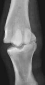 Animals are usually acutely and severely lame.
Elbow luxations can often be successfully reduced (returned to normal anatomy) and carry a good prognosis. For most other luxations, three months of casting or internal fixation is used to stabilize the limb. Arthrodesis is often attempted as the amount of joint damage is usually extensive, resulting in severe arthritis.
Joint luxations are relatively rare in large animal species. The exception is hip luxation in cattle.
Coxofemoral luxations in cattle
Cattle develop coxofemoral (hip) luxations with falls, by being ridden by other cattle when in heat, and subsequent to neurogenic injury (obturator paralysis). The hip can luxate craniodorsally or cranioventrally. Animals with a craniodorsal luxation can often stand; those with a cranioventral luxation cannot. The luxation can be identified by asymmetry of the bony prominences or via rectal palpation (cranioventral luxation). With a dorsal luxation, the limb is shortened, the hock turned inward and the toe drags
Cropped from https://www.youtube.com/watch?v=t-sR1bHhzKQ
Acute craniodorsal luxations are more successfully treated than more chronic or ventral luxations but only if there is no damage to the acetabulum. Traction is required. The cow is cast into recumbency with the affected limb uppermost. A rope is looped around the groin and secured to a fixed object (tree). Traction is applied to the limb to forcibly extend it, usually with a calf jack. Once the limb is extended, and the hock is rotated outward (upward) until the head of the femur slips back into the joint. If the joint is damaged, the hip will usually reluxate as soon as the traction is released.
Cranioventral luxations, luxations with trochanteric damage, or chronic luxations require arthrotomy and are very challenging to fix.
Tendon luxations
The most common tendon luxation in horses is luxation of the superficial digital flexor tendon. This occurs with damage to the retinaculum that holds the tendon to the calcaneus. As horses move, the SDF will visibly move as it runs over the hock. The luxation is usually on the lateral aspect and is associated with marked swelling over the point of the hock. The luxation may not be apparent in the standing animal. Horses are acutely and severely lame and may be very anxious when moving.
Treatment includes sedation and pain relief. If the tendon stays luxated, prolonged stall rest (4-6 months) is recommended. Limb movement may be minimized via a heavily padded bandage. A residual mechanical lameness (jerky motion) may remain after lameness improves. Horses may not be able to jump or race. Persistent medial luxations have a poorer prognosis for return to function.
Key Takeaways
Joint and tendon luxations lead to severe lameness. These are uncommon so call for advice.
Keep the animal calm and still while getting that advice.
Resources
Equid elbow luxation and subluxation: Anatomical review and case management considerations, Equine vet. Educ. (2022) 34 (7) 342-345
