Equine Lameness
Forelimb nerve anatomy
by Vic Cox
Anatomy
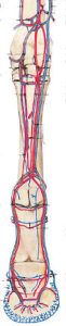
- The medial and lateral palmar nerves in the cannon region are adjacent to the deep digital flexor tendon. Much deeper are the medial and lateral palmar metacarpal nerves that lie on the palmar side of the metacarpus deep to the suspensory ligament. These nerves emerge at the distal ends of the splint bones and are sensory to the fetlock joint.
- At the fetlock the palmar nerves split to form the small dorsal
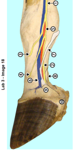
Superficial dissection of equine distal limb, freeze dried specimen with painted structures. 1, palmar nerve; 2, palmar digital nerve; 3, dorsal digital nerve; 4, intermediate digital nerve (not always present); 5, palmar digital artery; 6, palmar digital vein; 7, ligament of the ergot; 8, deep digital flexor tendon (DDFT); 9, collateral cartilage of P3; 10, bulb of the heel covered with periople. digital nerves and the larger palmar digital nerves which are the continuation of the palmar nerves. Note that the name is similar with digital inserted.
palmar digital nn. -> heel and navicular region
dorsal digital nn. -> toe and dorsum of digit
Nerve blocks
- The palmar digital nerves are the most commonly blocked nerves in the horse. They are blocked bilaterally in the midpastern for navicular and heel pain diagnosis and treatment. A horse with this block still knows where its toe is and therefore is safe to ride, but if blocked higher up so that the dorsal digital nerve is included, the horse is likely to stumble on an uneven surface.
- The palmar sesamoid block is at the level of the sesamoid bone and both the palmar and dorsal digital nerves are desensitized. Therefore, the entire digit will be desensitized with the possible exception of the proximal long pastern and the dorsal part of the fetlock joint.
- The low palmar (volar) block will block the palmar nerves below the communicating branch and also the palmar metacarpal nn. at the level of the distal ends of the splint bones.
- The high palmar (volar) block is performed above the communicating branch and blocks the same structures as the low palmar at a higher level. In turn, most of the suspensory ligament is blocked also.
- The distal blocks are usually done with the foot elevated but the upper two blocks can be done with the foot on the ground.
- Volar is a collective term meaning palmar or plantar. In most cases the nerve blocks are used in the forelimb but in occasional cases hind limb blocks may be needed.
Other clinical relevance
- While the palmar nerves follow the deep flexor tendon, the palmar digital nn. are attached to the palmar side of the digital arteries. The palmar digital nerve must be carefully dissected from the artery when a palmar digital neurectomy is performed
- The ligament of the ergot lies superficial to the palmar digital nerve and is sometimes mistaken for it during an attempted neurectomy. The ligament of the ergot crosses over the palmar digital nerve and digital vessels obliquely. While the nerve must be dissected off the digital artery, the ligament of the ergot is superficial and not attached to either of these structures.
