FA GI Topics
How to – Rumenotomy
Indications
Rumenotomy is indicated for vagal indigestion, hardware disease, rumen acidosis and some forms of choke. Rumenotomy allows exploration of the rumen, reticulum and parts of the omasum.
Relevant anatomy
The rumen is a large fermentation vat. The layers should be gas (dorsal), recent fiber, older fiber and fluid (ventral). The reticulum is sac off the dorsal aspect. The esophagus empties into the rumen from the dorsal cranial wall while the reticulum empties into the omasum on the medial wall just above the pillar separating the rumen and reticulum. The omasum and abomasum are often palpable through the rumen wall.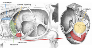
Preoperative management
Food restrictions: If possible the cow should be held off feed for 24 hours prior to rumenotomy.
NSAIDs/analgesics: Recommended preoperatively. Flunixin meglumine 1.1-2.2 mg/kg iv is standard.
Antibiotics:Recommended. If milking, ceftiofur 2.2 mg/kg im is standard. In beef or non-lactating cattle, other options do exist. Pencillin is often effective; however, remember that the label dose of procaine penicillin is ineffective!
Other: If possible, plan on rumen transfaunation.
Local blocks: A line block, inverted L or paravertebral are all reasonable options.
Position/preparation: See How to- Standing GI Surgery
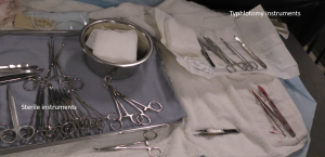
Surgery Supplies- Additional
- Extra sleeves and gloves
- Additional laceration or surgery pack is ideal
- Sterile saline for lavage
- Rumen board – optional
- Wound drape – optional
- 2 suture on a cutting needle for securing rumen
- 2-0 or 0 absorbable suture for closing rumenotomy
- Wet-dry vac -optional
Surgery Supplies – Standard
- Standard surgery pack
- Sterile sleeves for internal palpation
- Scalpel blade and handle
- 2 or 3 absorbable suture material for muscle closure
- 3+ nonabsorbable suture material for skin closure
Surgical procedure
Tacking sutures
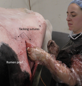
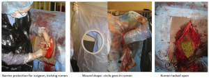
After exploratory, the rumen is tacked to the body wall to hold it in position. This is basically a modified set of stay sutures and is designed to minimize the work needed to hold the rumen in place while the sealing suture is performed. To tack:
- tack at approximately the 1:00 and 11:00 positions
- insert the needle (attached to #2 suture) through the skin away from the incision into the abdominal cavity
- grab a large bite of rumen away from the proposed rumen incision (keep the stay sutures out of the work area)
- drive the needle back through the skin near the original insertion and tie
Repeat until the rumen is held in position by the tacking sutures. Two -three sutures are typically sufficient.
Contamination management
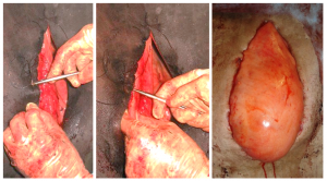
It is important to control contamination. This can be via use of a rumen board, rumen protector or by creating a water tight seal by use of an inverting suture pattern. The latter is the most effective.
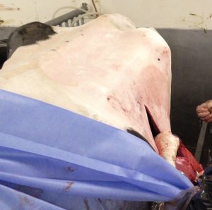
To create a water tight seal, secure the rumen to the skin using a continuous Cushing pattern of 2 suture on a cutting needle. Start the pattern with a basic simple knot and then start the Cushing pattern. Stop and tie suture as needed (at least once to prevent a purse string effect). When completed accurately, no suture should be visible. Any gaps should be closed with a mattress suture, especially at the ventral aspect. NOTE that the rumen should be poofed out a bit. However, too much poof leads to a poor match with the skin and more gaps due to the extra folds in the rumen.
To minimize contamination, separate rumenotomy instruments (scalpel, saline, gauzes, needle holders, suture, scissors) from closure instruments. Cover closure (clean) instruments with a towel to keep sterile and move them away from the area.
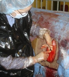
Consider adding protective gear to minimize lingering smells. Garbage bags are good protection. At this stage, the procedure is not a sterile one.
Rumenotomy
Incise the rumen. If available, place a wound protector. The plastic ring goes into the rumen and the plastic drape covers the rumen surface. If you don’t have a wound protector, another option would be to tack the rumen open or temporarily suture it open to minimize exposure of the serosal surface to sticky ingesta.
Remove ingesta along the path from incision to the reticulum for hardware disease. Remove all ingesta if the animal has rumen acidosis.
Explore the reticulum, esophageal opening and omasal opening. If the reticulum does not move, investigate the adhered area for penetrating foreign bodies. Palpate through the wall for any abscesses that need drainage. Hardware should be removed as should any extra magnets. One magnet should be left or placed. Rumen transfaunation is easiest at this stage.
If an abscess is identified, it can be drained into the rumen through the area adhered. Any incision that extends beyond that area will leak ingesta into the peritoneum. Note: Pus does not drain up hill well. And if you lose a scalpel blade, you have to find it. Tie it to your wrist before taking it into the rumen.
If contamination occurs or peritonitis identified, DO NOT lavage the abdomen. Cows can wall off infection if localized.
Rumen closure
The rumen drapes or shields are removed and the rumen surface cleaned. The rumenotomy incision is closed in two layers with an inverting pattern in the final layer. After the first layer is closed and the rumen surface cleaned, the garbage bags are removed from the surgeons, gloves changed and the clean instruments used for the second layer. After the second layer closure, the rumen is slowly released from the body wall so that it can be cleaned well prior to return to the abdominal cavity.
Body wall closure
Follow the guidelines for standing GI surgery. By this point, the drape may no longer be helpful.
Postoperative care
- Continue NSAIDs and antibiotics for at least 3 days
- Provide new rumen flora if possible
- Monitor for infection
Complications
- Lack of improvement
- Peritonitis
- Incisional infection
Videos
Resources
An incision made into the rumen for surgical exploration
traumatic reticuloperitonitis; foreign objects poke through the reticulum
suture pattern in which tissue edges are turned inward
