Female urogenital surgery
Milk flow issues- no milk flow
First calf heifers are occasionally presented with no milk flow from a teat, despite heavy milking in the other quarters. This can occur due to trauma, obstruction from infection or lactoliths or due to congenital anomalies.
Congenital anomalies can be a persistent membrane between the teat and the gland or lack of development of the teat cistern.
Diagnostics
Trauma, infection and milk stones are usually evident on physical examination.
A teat cannula can be passed up the teat and used as a gentle probe to evaluate the teat cistern.

A normal teat lining is smooth and does not obstruct passage of the teat cannula. If the teat cannula can be passed fully and easily, the most likely issue is a persistent membrane.
Ultrasound can be used to verify milk flow and occasionally can determine the cause of the obstruction. Fibrosis indicates prior damage and may be seen when calves suckle each other or when teats are stepped on .

Theloscopy (scoping the teat) can be used to evaluate prolapsed sphincters and other damage. This requires a needle scope and is not always available.
Therapy
Milk stones are uncommon and can be removed by forceful milking or grasping instruments. Trauma may respond, at least temporarily, with NSAID administration.
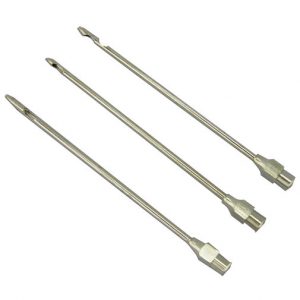
The membrane can be cut open through a thelotomy (incision in the teat) or via the teat sphincter. It is essential to avoid damage to the sphincter. Teat knives can cut through the membrane without stretching the sphincter. The membrane will tend to close over; frequent (q2h) milking for 3 days may keep it open. Using a teat implant is not advisable as these cause severe damage to the teat when the animal lies down. Alternatively, the quarter can be allowed to dry off. Total lactation amounts will be only slightly lowered.
A female bovid that has given birth to her first calf. This makes little sense since "heifer" refers to an immature female bovid but one that has maintained a pregnancy is obviously mature.

