Female urogenital surgery
How to – Minchev pexy
Indications
The Minchev technique is used for retention of vaginal prolapses particularly when animals will not be observed for parturition and when continued retention is likely (eg embryo flush cows).
Relevant anatomy
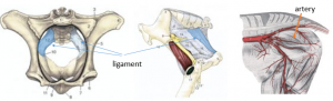
The suture is passed from the vaginal vault through the sacrosciatic ligament. The rectum and internal iliac artery should be avoided. The artery runs horizontally across the pelvis and the pulse is palpable.
Preoperative management
Food restrictions: NA
NSAIDs/analgesics: Perioperative NSAIDs are recommended.
Antibiotics: Preoperative antibiotics are recommended.
Local blocks: Epidural, pudendal and/or line block
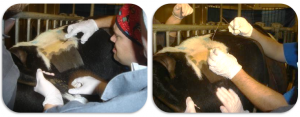
Position/preparation: The patient is restrained standing. The surgeon is gloved. The vagina is cleaned as is the skin over the gluteal muscles. An assistant with a rectal sleeve can be used to move the rectum out of the line of fire.
Surgery Supplies:
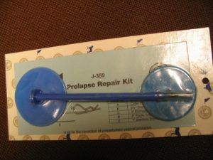
- Straight needle (strong, at least 3″ long), Buhner needle (ouch), S needle or Minchev kit
- Umbilical tape or large nonabsorbable suture
- Buttons, inserts from ligapak, gauze rolls or stents
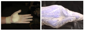
- Gauze (padding for glove) or thimble- optional
Surgical procedure
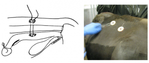
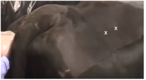
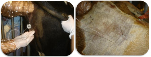
The surgeon palpates vaginally to find the internal iliac artery and determine placement of two buttons. These should be placed as close to the cervix as possible. The surgeon can push on the vaginal wall and see the exit site on the outside of the cow.
The needle is inserted vaginally and exited out the vaginal wall at 1-2:00, avoiding the artery. Manipulation is minimal due to the size of the instruments. The prolapse device has a locking pin and buttons. It is self-retaining after the stylet is removed and the pin inserted.
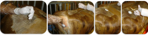
Buhner needle
Once the Buhner needle is exited, umbilical tape is threaded through it and it pulled back into the vagina, bringing the tape through. The procedure is repeated to create a horizontal mattress. The tape is threaded through a stent, button or around gauze both externally and internally to prevent suture pull through.
A second pexy is repeated on the same side to strengthen the adhesion without constricting the rectum.
Straight needle or S needle
With the straight needle, a long piece of suture is threaded on the needle. The needle is inserted into the vaginal wall at the desired location.
The surgeon pushes the needle through (a thimble inside the glove or a gauze padded palm make this less painful if using a straight needle) and an assistant grabs it as it exits.
One strand of the suture is pulled to the outside and held. The internal strand is threaded through a button or stent and the needle replaced on that strand. The procedure is repeated close to the first puncture.
Both ends are tied over a second button externally.
A second pexy is repeated on the same side to strengthen the adhesion without constricting the rectum.
Rectal palpation should be used to verify no suture has entered the lumen.
Postoperative care
- Sutures should be left in place through parturition or at least for 3 weeks if the animal is not pregnant.
Complications
Infection is common but rarely leads to an abscess. Suture that enters the lumen of the rectum should be removed.
Prolapse is still possible, particularly if the pexy isn’t far enough cranial, if the sutures tear out or if just bad luck.
Videos
Resources
Using the prolapse kit
