FA GI Diagnostics & GI Surgery Principles
How to – Bovine Abdominal Exploratory- Left
Indications
Left flank exploratories are less common but are useful when the animal is showing signs of vagal indigestion or other left sided abnormalities. Anytime a procedure is performed on the left (Csection, rumenotomy) it should be accompanied by a full exploratory.
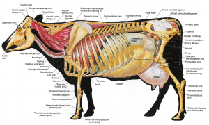
Relevant anatomy
The rumen takes up considerable space. How much of the intestinal structures are palpable depends on the size of the cow, arm length of the surgeon and the amount of intestinal fill.
Preoperative management
Food restrictions: NA
NSAIDs/analgesics: Recommended preoperatively. Flunixin meglumine 1.1-2.2 mg/kg iv is standard.
Antibiotics: Recommended. As cows can wall off infection, there can be surprises inside. If milking, ceftiofur 2.2 mg/kg im is standard. In beef or non-lactating cattle, other options do exist. Remember: the label dose of procaine penicillin is ineffective!
Local blocks: A line block, inverted L or paravertebral are all reasonable options.
Position/preparation: See How to- Standing GI Surgery
Surgery Supplies:
- Standard surgery pack
- Sterile sleeves for internal palpation
- Scalpel blade and handle
- 2 or 3 absorbable suture material for muscle closure
- 3+ nonabsorbable suture material for skin closure
- Biopsy instruments and formalin- optional
- DA simplex – long flexible tubing attached to a 10ga needle for viscus decompression -optional
Surgical procedure
Follow the guidelines for standing GI surgery.
Place a sterile sleeve on your right arm (regardless of whether left or right handed). If it is large for your hand, add a sterile glove over the top to improve your palpation skills. Generally this is one size larger than your normal glove size. **REMOVE ANY HEMOSTATS USED FOR BLEEDERS**. Finding these in the belly is somewhat challenging.
Exploration should be done from clean to potentially (or known) contaminated regions. In cattle, the riskiest areas are by the reticulum (hardware disease), liver (abscesses) and abomasum (perforated ulcers). As all of these are cranioventral, we typically start caudodorsal and to the right, followed by caudoventral, craniodorsal and finally, cranioventral.
- Reach caudally and curve your right arm so that you can check the intestinal fill on the right side.
- Return to the pelvis. Evaluate the structures in this region:
-
- bladder – she will urinate
- sublumbar lymph nodes
- uterine – check involution
- ovaries – evaluate for normal size and structure
-
- Evaluate the rumen pack
- Normal fill and layers?
- Evaluate the ventral abdominal floor. Pass your hand along the ventral abdomen. It should pass easily without obstruction.
- Evaluate the cranial abdomen
- Palpate the reticulum – is it freely movable? Adhered?
- If no issues, reach across to the abomasum.
- Normal fill and position?
- Evaluate the diaphragm and spleen
- Diaphragm should be intact with no herniation or defect.
- Spleen should be smooth, not nodular
Adhesions should NOT be broken down. They are usually attempting to wall something off.
Closure : Follow the guidelines for standing GI surgery.
Postoperative care
Complications
Videos
Resources
traumatic reticuloperitonitis; foreign objects poke through the reticulum
