LA Respiratory Issues
Pharyngeal disorders
The pharyngeal region includes the soft palate, extensions of the palate into the walls and roof of the pharynx, openings into the guttural pouches and to the larynx. Pharyngeal dysfunction is an important cause of poor performance and noise associated with exercise. The muscles of the pharynx are essential to maintaining an open airway despite high negative pressures during inspiration.
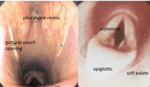
Common disorders
Cleft palate
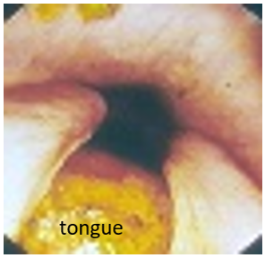 Cleft palates occur as congenital anomalies in most species. The earliest clinical sign is usually milk drainage from the nostrils. Careful digital evaluation of the palate should be performed in all neonates to ensure the palate is complete. Surgical repair is challenging and success depends on how much of a defect is present and whether or not aspiration pneumonia develops. If treatment is an option, affected animals should be hospitalized as soon as possible to minimize the risk of aspiration through feeding tube placement and antibiotic therapy. The image above is an endoscopic view of a cleft palate as seen from the nasal passages.
Clinical signs – milk nasal discharge, cough
Pharyngeal lymphoid hyperplasia/ Pharyngitis
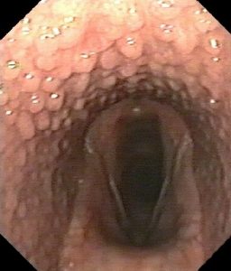
The lymphoid tissue in the pharynx can become inflamed in young horses leading to numerous bumps visible endoscopically. Pharyngeal lymphoid hyperplasia (PLH) may lead to symptoms of a sore throat but is often identified incidentally. This is most common in race horses being “scoped” to figure out why they aren’t running as fast as the trainer/owner had hoped. There is no noise associated with PLH unless the inflammation leads to other problems. Treatment is not usually required but throat sprays containing an anti-inflammatory agent may help.
Clinical signs – may have decreased appetite or dysphagia but this is rare
Dorsal displacement of the soft palate
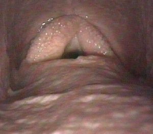
In the horse, the soft palate merges into the walls of the pharynx, totally separating the oral cavity from the nasal cavity. The larynx connects with the nasal passage through an opening in the soft palate. When the horse swallows, the palate moves up to allow food to access the esophagus (lives dorsal to the larynx). With DDSP, the palate moves up during exercise, blocking part of the airflow. Diagnosis is through exercise endoscopy. The most common presentation is a horse that “stops” abruptly during intense exercise. A gurgling noise is often associated. The stopping is due to the displacement of the palate over the airway, preventing sufficient air and oxygen flow. The gurgling is due to vibration of the palate on exhalation. Multiple causes of DDSP exist, leading to various treatment options. As one of the main contributing factors is inflammation in the throat or guttural pouch inflammation, NSAIDs and steroid throat sprays are typically the first line of treatment. If anti-inflammatory treatment does not help, the preferred surgery is a “tie-forward” procedure to keep the larynx positioned within the ostium (opening in the soft palate). Tongue-ties, while not supported by clinical trials, are common in racing horses and are designed to prevent DDSP by preventing swallowing during racing.
Clinical signs – noise with exercise, sudden stopping while working
Pharyngeal collapse
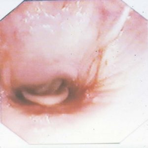
Pharyngeal collapse results from the large negative airway pressures and is a common cause of poor performance. Treatment is nonspecific and often not very helpful. Diagnosis is through endoscopy during exercise. Any inflammation and other disorders should be treated. The prognosis is poor for most performance horses.
Note : pharyngeal collapse can occur in young animals with muscle disorders (lack of tone in the pharyngeal muscles). Pharyngeal narrowing can also occur due to pressure from outside structures (full guttural pouch, enlarged retropharyngeal lymph nodes). This type of narrowing is visible on standing endoscopy and lateral head radiographs.
Clinical signs – noise with exercise, poor performance
Rostral displacement of the palatopharyngeal arch
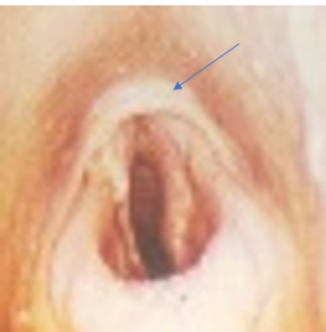 The palatopharyngeal arch borders the opening to the larynx. When rostral displacement occurs, the opening to the larynx is partially obstructed. This is a congenital lesion leading to impaired laryngeal function (required for performance) and swallowing. The arytenoids can’t abduct (open) properly due to the restrictive band. Many of these horses have 4-BAD – 4th branchial arch defect. Treatment is often unrewarding.
Clinical signs: poor performance, noise with exercise
Pharyngeal trauma
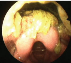
Pharyngeal trauma is relatively common in cattle, often due to medication administration with bolus guns. The bolus can be pushed through the oropharynx, causing cellulitis. Cellulitis in this area can lead to dysphagia, dyspnea and mediastinitis. Diagnosis often requires radiographs and endoscopy; oral palpation can be useful in larger patients. To treat, the foreign body is removed, any abscesses are drained surgically and the cellulitis treated with systemic antibiotics. A rumen fistula may be needed for feeding and a tracheotomy needed for breathing in more severe cases. Due to the location, drainage of the abscesses can be tricky and is best done at a referral hospital, if possible.
Clinical signs – malodorous breath, anorexia, fever, dysphagia
Resources
Respiratory distress in the adult and foal, VCNA (2021) 37:311-325
Upper airway conditions affecting the equine athlete, VCNA: Equine Practice, August 2018, Vol.34(2), pp.427-441
Update on diseases and treatment of the pharynx, VCNA: Equine Practice, April 2015, Vol.31(1), pp.1-1
Surgery of the Equine Upper respiratory tract : focus on dynamic disorders. Proceedings, 2013
Respiratory Surgery, Vet Clin Food Anim 32 (2016) 593-615
Staphylectomy in nonbrachycephalic dogs, CVJ / VOL 64 / AUGUST 2023- I suspect we will find this isn’t quite as simple as we think right now – different anatomy, more complications

