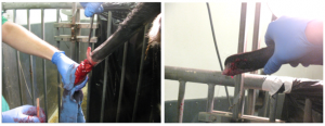
FA youngstock processing
How to – Tail Dock – Adult
Indications
Tail docking in adult animals should be restricted to cases of trauma or infection.
Relevant anatomy
The tail is comprised of vertebrae, vertebral spaces, coccygeal muscles and vessels on either side. Disarticulation should be between vertebrae.
Preoperative management
Food restrictions:NA
NSAIDs/analgesics: Perioperative NSAIDs are recommended
Antibiotics: NA
Tetanus prophylaxis is recommended in horses
Local blocks: Epidural or ring block
Position/preparation: The patient is kept standing. Surgeon wears gloves. The area is clipped and prepped.
Surgery Supplies:
- Scalpel and handle
- Mosquito hemostats
- Mayo scissors
- Needle holders
- Suture scissors
- 3-0 absorbable (vessels)
- 2-0 or 0 suture, cutting needle (skin)
- Tourniquet – optional
Surgical procedure
A tourniquet may be applied proximally but usually isn’t necessary.
The appropriate vertebral space is identified. Semilunar skin incisions are made dorsally and ventrally with both flaps extending beyond the point of disarticulation. The dorsal flap should extend further than the ventral flap. The flaps are undermined and retracted cranially. The vessels are ligated with 3-0 suture and muscles transected. The coccygeal vertebrae are disarticulated. The dorsal flap is folded over the end and sutured to the ventral flap.

Postoperative care
- Keep area clean and dry.
- Suture removal in 10-14 days
Complications
Dehiscence and infection are possible. Second intention healing is recommended if either occur.
Videos
Resources
See Small Animal Surgery Textbook, Theresa Welch Fossum, Ch 16 Surgery of the Integument
P Aubry. Routine Surgical Procedures in Dairy Cattle Under Field Conditions: Abomasal Surgery, Dehorning, and Tail Docking. Vet Clin Food Anim 21 (2005) 55–72

