FA GI Diagnostics & GI Surgery Principles
How to – Bovine Abdominal Exploratory- Right
Indications
An abdominal exploratory is a relatively cheap and effective diagnostic tool both in the field and in the hospital. Since most cows will not be shipped to a referral center, many diagnostics are not available. Lab-work typically needs to be run at the clinic or shipped off. Ultrasound is a powerful tool and can be utilized in the field. In most cases with an abnormal ultrasound, exploratory surgery is indicated. If ultrasound is negative, the next useful tool is exploratory laparotomy.
Relevant anatomy
The right flank is the preferred approach to an exploratory. Due to the rumen, ventral incisions are useful only for direct evaluation of structures in the abomasal area. From the right flank, most structures are palpable if not visible.


Preoperative management
Food restrictions: NA
NSAIDs/analgesics: Recommended preoperatively. Flunixin meglumine 1.1-2.2 mg/kg iv is standard.
Antibiotics: Recommended. As cows can wall off infection, there can be surprises inside. If milking, ceftiofur 2.2 mg/kg im is standard. In beef or non-lactating cattle, other options do exist. Remember: the label dose of procaine penicillin is ineffective!
Local blocks: A line block, inverted L or paravertebral are all reasonable options.
Position/preparation: See How to- Standing GI Surgery
Surgery Supplies:
- Standard surgery pack
- Sterile sleeves for internal palpation
- Scalpel blade and handle
- 2 or 3 absorbable suture material for muscle closure
- 3+ nonabsorbable suture material for skin closure
- Biopsy instruments and formalin- optional
- DA simplex – long flexible tubing attached to a 10ga needle for viscus decompression -optional
Surgical procedure
Follow the guidelines for standing GI surgery.
Place a sterile sleeve on your left arm (regardless of whether left or right handed). If it is large for your hand, add a sterile glove over the top to improve your palpation skills. Generally this is one size larger than your normal glove size. **REMOVE ANY HEMOSTATS USED FOR BLEEDERS**. Finding these in the belly is somewhat challenging.
Exploration should be done from clean to potentially (or known) contaminated regions. In cattle, the riskiest areas are by the reticulum (hardware disease), liver (abscesses) and abomasum (perforated ulcers). As all of these are cranioventral, we typically start caudodorsal and to the left, followed by caudo ventral, craniodorsal and finally, cranioventral.
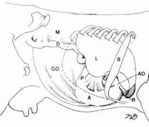
The omental sling will be in the palpation path unless pulled out of position by a late term pregnancy. In most cases, the duodenum and sling will be the only things visible in the incision (Fig 1). If there is a distended structure, it can be the abomasum (RDA or abomasal torsion) or gas in the omental bursa. Gas in the omental bursa develops after abdominal surgery or with a ruptured abomasal ulcer.
- Reach caudally and curve your left arm so that you can check the rumen and the left side. Palpate forward along the left body wall. Check the rumen gas cap and rumen pack. The rumen should have a small gas cap and be easily indentable.
- An LDA will palpate as a second rumen gas cap.
- Return to the pelvis. Evaluate the structures in this region:
- bladder – she will urinate
- sublumbar lymph nodes
- uterine – check involution
- ovaries – evaluate for normal size and structure
- Evaluate the caudal intestines. The intestines should be fairly empty and located in the back 1/3 of the abdomen.
- The intestines cannot be traced from duodenum to cecum
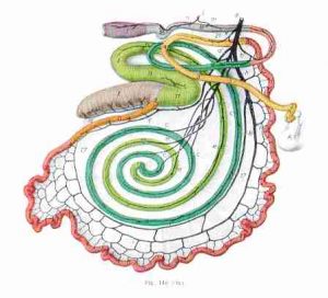
- If the intestines need to be evaluated, exteriorize the cecum (a blind-ended sac with no bands). The spiral colon is connected on the dorsal surface and the ileum enters on the ventral surface. (Fig 3)
- Evaluate the kidneys. Reach forward on the inner aspect of the sling. The kidneys are both on the right side of midline, lobulated and should be nonpainful.
- Evaluate the ventral abdominal floor. Pass your hand along the ventral abdomen. It should pass easily without obstruction.
- Return to the outer surface of the sling and palpate the cranial abdomen.
- Evaluate the liver
- Edges should be sharp. Rounded edges indicate likely fatty liver.
- The gall bladder will be distended in cattle that are anorexic.
- Evaluate the ascending duodenum.
- It should not be distended. Normal attachments connect it to the liver.
- Evaluate the abomasum
- The abomasum can be identified by a mucosal slip.
- The abdomen should be mostly empty and should be located on the right side of midline in the ventral abdomen
- The pylorus is muscular but should be evenly muscled and a lumen should be palpable.
- Evaluate the omasum
- This is a round firm basketball medial to the abomasum
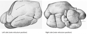
- Evaluate the reticulum (Fig 2)
- To find the reticulum, pass your hand down along the body wall, palm side down. Aim for the ventral midline about 6-12″ forward from the incision (NOT up by the tonsils). Turn your hand palm side up. The reticulum will usually be in your palm. The reticulum is a mostly empty sack that should be easily movable. It often contains sand. The honeycomb pattern is variably distinct.
- Evaluate the diaphragm
- It should be intact with no herniation or defect.
- Note: When performing an exploratory on a cow who has recently calved, make sure to start caudodorsally to avoid contamination risks in the cranioventral abdomen
Closure : Follow the guidelines for standing GI surgery.
Postoperative care
Complications
Videos
We try not to break down adhesions in cattle; however, if she has had an omentopexy and this is impeding exploration, it may be necessary.
Resources
traumatic reticuloperitonitis; foreign objects poke through the reticulum
tacking the omentum in place to prevent movement of the abomasum

