Equine Lameness
Gait abnormalities
These conditions are not always painful so will not block out. Being able to identify them will save you time and trouble!
Sweeney
Etiology: Atrophy of the shoulder muscles occurs. This allows the shoulder to luxate since there are no collateral ligaments of the shoulder joint. Muscles such as supra- /infraspinatus, subscapularis are so called “active collateral ligaments” but are not as restrictive as true ligaments. In the past, sweeney was associated with cart horses and pressure placed on the suprascapular nerve by the cart braces. Now it is most often associated with trauma to the shoulder: eg a kick or fracture that damages the nerve or when a horse runs into a gate or fence post.
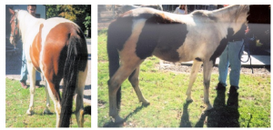
Clinical signs : Shoulder joint moves outward from the scapula.
Treatment : rest, anti-inflammatory drugs, surgery to decompress the nerve
Surgery is not usually performed until after 3 months of rest and waiting to see if the muscles will regain tone
Prognosis : guarded to poor for return to normal function
Upward fixation of the patella
Etiology : lack of tone in the quadriceps, allowing the patella to lock in place. This is most common in young horses and those that have been out of training for awhile and have quadriceps muscle atrophy
Clinical signs : stiff extended leg, particularly when the horse is backed up. May be bilateral . When the stifle “unlocks” horse moves forward normally. You may be able to manually move patella into the locked position fairly readily
Treatment: the old treatment was to cut the medial patellar ligament, thereby preventing the locking. However, normal horses that have this surgery have a strong tendency to develop arthritis in the stifle joint. Now we recommend exercise to strengthen the quads: hill work, over cavalettis (wood poles on the ground), etc. Other treatments include tendon splitting, irritants injected around the medial patellar tendon to tighten it up, and hormone injections to change the amount of ligamentous/tendinous relaxation.
Prognosis : good if no other problems with the stifle joint
Locking Stifle/Upward Fixation of the Patella in a horse. (Kaltura version w/ captions)
Fibrotic myopathy
Etiology : trauma to the semitendinosus muscles (injection, injury)
Clinical signs: restricted forward motion to the hind limb action at a walk. Limb moves forward only so far and then is slapped to the ground due to the restricted muscle action. The gait abnormality is not apparent at faster gaits
Treatment : the old treatment was to resect the scar tissue but this made more scar tissue. Now most people either ignore it, transect the tendon of insertion of the semitendinosus muscle, or cut some of the restrictive areas under local anesthesia in the standing horse.
Prognosis: there are complications associated with the treatment; if it is mild, it may be best to avoid surgery
Peroneus Tertius Rupture (Kaltura version w/ captions)
Peroneus (fibularis) tertius rupture
Etiology: overextension of the hind limb such as when it is caught in or under something (eg trailer). It may also be iatrogenic during recovery from anesthesia when the leg is in a full limb cast. This occurs because the cast makes the hock more immobile than the stifle joint.
Clinical signs: loss of reciprocal apparatus — the hock can be extended when the stifle is flexed and the hock does not flex much when the horse is moving (straight leg)
Anatomy note: The peroneus tertius and superficial flexor tendon work together as the reciprocal apparatus to efficiently move the limb as a unit. Add in the patellar ligaments locking on the femur and the horse can sleep standing up. If the horse doesn’t have good quadriceps muscles, it can’t unlock the stifle as easily so you get the hitch seen with locking patellas. Lose the peroneus tertius and you can extend the hock while flexing the stifle (ouch)
Treatment: rest and anti-inflammatory drugs
Prognosis: good
Peroneus Tertius Rupture (Kaltura version w/ captions)
Stringhalt
Etiology: thought to be neuronal dysfunction or adhesions around the lateral digital extensor tendon; also due to a plant toxin in Australia
Clinical signs: over flexion of the limb during the forward phase of the stride –the hind limb is pulled up in an exaggerated motion and may even hit the ventral abdomen. May be bilateral.
Treatment for pasture associated forms: Most horses will recover with time; it can take 6-24 months. Phenytoin (15 mg/kg BW every 12-24h) may help but blood levels should be monitored. Lateral digital extensor myotenectomy is not recommended.
Treatment for non-pasture associated forms: Lateral digital myotenectomy may help some forms but the gait abnormality may also recur.
Prognosis : Good for pasture associated forms. Guarded for other forms.
Shivers
Etiology : neuromuscular disease that likely involves an abnormality in the feedback loop between afferent and efferent nerve fibers
Clinical signs : involuntary jerky flexion of the pelvic limb (and testicles) as well as extension of the tail: leg is held off the ground in a flexed position and muscles of hind limb and tail may quiver. Mild cases may be intermittent. Generally noted when horse is backed, turned, or made to step over an object. Most common in draft breeds.
Treatment : no specific treatment. Treat any other abnormalities such as lameness or polysaccharide storage myopathy as these will make it worse. Supplement with Vitamin E if levels are low. Horses may do better with regular exercise, limited stall rest, good nutrition and low stress.
Sedation may be needed to permit hoof trimming in the hindlimbs.
Prognosis : slowly progressive; overall prognosis is poor
It is important to determine that the signs aren’t caused by PSSM (polysaccharide storage myopathy) as that disease is very treatable
Gastrocnemius rupture
Etiology : Foals may develop gastrocnemius rupture during dystocia and with assisted delivery. Rupture of the gastrocnemius muscle occurs when the stifle is forced into overextension while the tarsus is flexed. Adult horses may develop gastrocnemius rupture during exercise (in humans, this is generally with twisting; in horses with strenuous stopping).
Clinical signs: Dropped tarsus with extended stifle. inability to straighten the hindlimb. Mild to severe swelling in the upper limb. If the rupture is complete, the horse is unable to bear weight on the affected limb. Bilateral hindlimb gastrocnemius ruptures will result in recumbency due to the inability to stand. Ultrasound, radiographs (calcified injury area) and CT have been used to confirm incomplete ruptures.
Treatment : stall rest, fixation of the affected limb by a full limb cast and/or Robert Jones bandage and administration of anti-inflammatory agents have been used with varying success. Lack of stabilization of the limb makes casts challenging as rub sores are almost inevitable. Affected foals have been reported to return to full function without bandaging or splinting. Intensive supportive care is needed to ensure the foal can stand to nurse. The contralateral limb is at risk of developing angular limb deformities (foals) or laminitis (adults) due to increased weight bearing.
Prognosis : fair if incomplete, guarded to grave if complete rupture
Resources
Shivering and stringhalt in horses, The Veterinary Journal 282 (2022) 105829
