Large animal wounds
Cellulitis
 Cellulitis is disseminated infection under the skin – the bacteria grow in the tissue planes rather than in an abscess capsule.
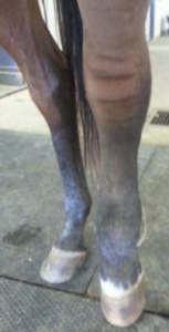
The affected area becomes warm, swollen and painful. Animals will be febrile and have changes in white blood cell counts.
These horses are very lame; it can be difficult to rule out a septic joint. Do not tap the joint as that WILL take bacteria into the joint. Ultrasound can be difficult as the pressure of the procedure really hurts. Radiographs may show gas in the joint if it is infected but infection may be present without gas. Most times we treat for cellulitis and warn clients that there may be more. Most times it is just cellulitis. [Future – researchers are working on enzymatic markers of synovial infection; this type of stall side test would make joint infections much easier to identify!].
Differentials
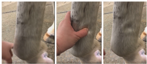
Differentials include edema and lymphangitis. Edema occurs due to leakage of fluid from vessels into the extravascular space. Edema reflects changes in Starling’s forces. Most often, edema is related to low protein in the blood. This means the fluid in the bloodstream flows down the concentration gradient into the tissues. Alternatively, the hydrostatic pressures in the vascular system are higher than normal and shove fluid out. Edema results in a thickened limb that is cool. When a thumb is pressed into the limb, a depression will remain for a few seconds (pitting edema- video link).
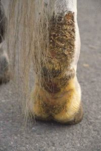
Lymphangitis is inflammation of the lymph system that interferes with movement of fluid out of the limb. It can develop secondary to dermatitis or systemic infections (eg Corynebacterium pseudotuberculosis). Lymphangitis will also lead to edema and is often associated with bacterial infections that block the lymphatic system. Ulcerative skin lesions may develop.
Cellulitis and lymphangitis often lead to secondary edema due to compression of the lymphatics and capillaries. That compression impedes venous return and the removal of normal fluid. If a horse has edema secondary to heart failure or low protein, edema should be evident in other regions of the body; not just the limb.
Therapy for cellulitis includes systemic antibiotics and NSAIDs. When open wounds are cultured, Staphylococcus aureus is a common finding. Therapy often starts with trimethoprim sulfadiazene or doxycycline; however, oral antibiotics are not always sufficient. Intravenous regional antibiotics (penicillin/gentamicin or ceftiofur) may be useful to increase drug concentration in the affected area. Limbs are also bandaged to remove swelling and decrease pain.
If left swollen, limbs will develop problems with lymphatic and capillary flow. Many affected horses will be prone to limb edema with stall rest.
Complications include contralateral limb laminitis, dermal necrosis, persistent lameness, septic tendonitis/desmitis and even hoof capsule avulsion (falling off).
Key Takeaways
If the limb is swollen, hot and painful, think cellulitis. Treat with antibiotics and firm wraps to push the fluid out of the limb.
Resources
A cross‐sectional survey of the diagnosis and treatment of distal limb cellulitis in horses by veterinary surgeons in the United Kingdom. April 2021Equine Veterinary Education 34(6)- good images and hints on diagnostics and therapeutics in a field setting
Treatment and outcome of eight horses with limb cellulitis and septic tendonitis or desmitis. Veterinary Surgery. 2021;50:1542–1552.
inflammation of a ligament
