FA GI Topics
Cecal dilation
Cecal dilation is a relatively common disorder in postpartum dairy cows.
Signalment
Signalment predisposition is the same as LDAs– dairy cattle in the first 6-8 weeks after parturition, particularly those with other disorders. In the first few days after parturition, cecal dilation is seen in those cows with hypocalcemia. [Hypocalcemia occurs due to the heavy shift of calcium to milk production.].
Clinical signs
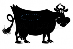
Right sided ping, extending horizontally below the transverse processes. The ping may extend up to 3 feet in length. Cows with hypocalcemia often have cold ears and slow pupillary light responses. Cattle with hypocalcemia may have overall gut stasis and also have signs of rumen bloat. The ping may resemble an RDA except it is more horizontal and extends further caudally.

Rectal examination
A large viscus (long balloon) can be palpated in the pelvic inlet to the right of midline
Treatment
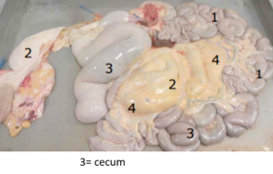
While cattle may respond to medical therapy (calcium supplementation), surgery can help by relieving the distension. The cecum is approached via right paralumbar fossa incision. In most cases, the distended cecum is readily identified with palpation on the inner aspect of the omental sling. The bovine cecum is just a plain sac- no bands or sacculations.

The cecum can be exteriorized for gas decompression (10-14ga needle inserted through a cruciate suture) or via typhlotomy (incision at the tip of the cecum).
The cecum may twist (cecal volvulus), causing ischemic necrosis. These cattle may show signs of colic. If cecal necrosis is identified, the necrotic portion of the cecum can be removed (typhlectomy) via standing surgery.
Resources
Surgery of the bovine large intestine, VCNA 2008
Clinical findings and treatment in cows with caecal dilatation, Vet Record 2012

