Swine, SRC, and poultry musculoskeletal disorders
Camelid bone, muscle and tendon disorders
Anatomy
- The superficial digital flexor tendon has a direct fascial connection to the proximal suspensory ligament (so issues with one tend to impact both)
- More in Chapter 58 – Musculoskeletal Surgery of Llama and Alpaca Care
- Smaller camelids can manage with 3 limbs so amputation is a feasible option. Similarly, postoperative care and fracture management are easier due to the ability to use slings and various splints. A light weight body also means camelids can be treated with implants designed for dogs, horses or humans; hence, a wider variety of options exists for repair of injuries or abnormalities.
Flexural deformities
Flexural deformities are sometimes encountered and are treated similarly to foals. In these cases, the tendons are “tight” and restrict the limb motion so that it can’t straighten. In camelids, we can be more aggressive with surgery due to the limited athletic requirements.
Suspensory degeneration /Fetlock hyperextension
Camelids can develop “dropped fetlocks” or bilateral conformational abnormalities due to what is believed to be suspensory degeneration. This syndrome has been associated with mineral deficiencies, abnormal proteoglycan levels in the suspensory ligament and genetic predisposition. We see similar lesions in horses. In both species, no effective therapy exists and prognosis is poor.
Unilateral cases of dropped fetlocks can occur with excessive weight bearing and are more likely to be due to suspensory damage rather than degeneration.
Juvenile osseous sequestration
While horses and cattle develop bony sequestration after wounding, many of the sequestra diagnosed in camelids do not have open wounds associated. This form of sequestration is most common in young camelids and it more closely resembles a syndrome seen in children in which bacterial emboli seed bone, leading to bone ischemia.
Long bones of the appendicular skeleton seem to be most affected. Animals present with signs of heat, pain, local swelling and lameness. Diagnosis is made radiographically.
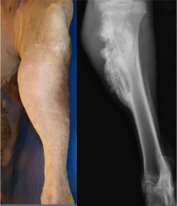
Sequestrectomy (removal of the sequestrum) usually results in a good prognosis as long as enough supporting bone remains to prevent fracture. Culturing the bone is recommended.
Fractures
see orthopedic emergencies chapter
Congenital lesions
Camelids have significant numbers of congenital lesions. Orthopedic lesions include wry nose, polydactyly (extra toes), syndactyly (fused toes), patellar luxation, flexural deformities and rotated talus bones. The rotated talus bones are associated with an abnormal gait.
Key Takeaways
Camelids do not get too many lameness issues.
Be aware of
- suspensory degeneration leading to fetlock hyperextension
- juvenile osseous sequestration unrelated to open wounds
- patellar luxations, potentially related to early castration
Camelids can be treated much more like dogs in terms of joint luxations, etc. Their light bodies mean they are able to ambulate on three limbs and we have more repair options.
Resources
Pain management in small ruminants and camelids: Analgesic agents.Vet Clin North Am Food Anim Pract . 2021 Mar;37(1):1-16.
Llama and Alpaca Care. Medicine, Surgery, Reproduction, Nutrition, and Herd Health. Elsevier, 2014.
pieces of dead bone that cause persistent draining tracts as the body tries to liquefy them
