FA GI Topics
Other abomasal disorders
Abomasal bloat
Young calves, lambs and goat kids can get abomasitis. Depression, abdominal distension and occasional colic signs can develop rapidly, usually in animals less than 3 weeks of age. The abomasum becomes gas filled and inflamed; at necropsy, it often contains foul smelling clots of milk and bile reflux from the small intestine. Hemorrhage into the lumen and/or perforating ulcers may be evident. The disorder is highly fatal. The etiology is not well understood but may relate to Clostridial infections and gas production connected to the highly fermentable foodstuffs.
Differential – rumen bloat or abomasal displacement
Risk factors seem to be related to changes or inconsistencies in feeding practices. These include interruption of normal nursing patterns in beef calves due to weather etc. In dairy calves, it may be related to feeding of large volumes of milk, cool milk temperature and poor milk hygiene.
Early intervention is crucial.
- Do not feed the calf anything that can be fermented (no milk, no glucose containing electrolyte solutions)
- SQ penicillin to treat abomasitis
- IV or SQ fluids
- Flunixin meglumine iv
- If needed, you can remove gas from the abomasum but the calf needs to be placed on its back to get the gas to float up
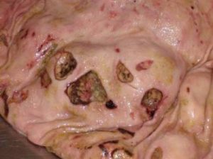
Abomasal ulcers
Abomasal ulcers may bleed or perforate. Surgery is not advisable when a cow has a bleeding ulcer. However, ulcers are commonly associated with abomasal displacement so surgery may be unavoidable.
A cow with a ruptured abomasal ulcer may show signs similar to hardware disease initially. Ruptured ulcers lead to localized peritonitis. Cattle are really good at walling off infections. In fact, they may go on to look perfectly normal. When you encounter an adhesion at surgery, LEAVE IT ALONE unless the cow will not survive without you breaking it down. You are likely going to open up an abscess. Similarly, we don’t flush the abdominal cavity in ruminants with peritonitis. All you are doing is dispersing the infection so the cow has to wall off multiple areas.
If the cow has a displaced abomasum along with an ulcer, the ulcer and resultant peritonitis may keep the abomasum from floating as high as it normally would. If you have low ping, worry about a ruptured ulcer!
At surgery, if the omental bursa is gas distended, assume you have a ruptured abomasal ulcer on the lesser curvature. Due to the omental leaves, the gas is trapped in the bursa.
When an ulcer is suspected, avoid tugging on the abomasum more than necessary. A right paramedian approach is recommended, as this will provide the best exposure to the abomasum and avoid extra manipulation. However, if the cow is anemic, the dorsal recumbency can be dangerous. If you are working from a right flank approach, do not pull the abomasum up any higher than necessary. Tack it lower than you usually would and let her recover.
Note: midlactation cattle with signs of abomasal ulceration should be checked for lymphosarcoma
Abomasal impaction
Impactions are not common but may be seen with changes in feedstuff, after abomasal volvulus and with pyloric lymphosarcoma. Cattle with lymphosarcoma or with impaction after volvulus should be culled.

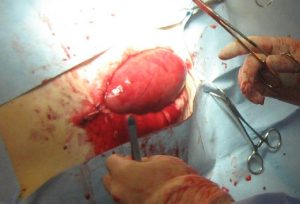
Abomasotomy is performed for abomasal impactions due to a change in feedstuff. It is performed with the animal sedated in left lateral recumbency.

If the animal has rumen distension, a rumenotomy should be performed first to empty the rumen. Otherwise, the rumen distension may lead to significant ventilation issues.
Resources
- Right flank laparotomy and abomasotomy for removal of a phytobezoar
in a standing cow. CVJ 51(7):761-3
traumatic reticuloperitonitis; foreign objects poke through the reticulum
blockage or partial blockage of the intestinal lumen by feedstuff
remove from the herd, usually by shipping to slaughter but animals could be euthanized or slaughtered on the farm.

