LA umbilical disorders
Umbilical hernias
Umbilical hernias- overview
Definitions
Abdominal hernia definition: protrusion of one or more abdominal organs through a congenital or acquired opening in the abdominal wall.
Parts of a hernia: the contents of the hernia exit the cavity they are normally contained through a defect referred to as the hernial ring and enter a hernial sac.
Congenital hernia: present at birth.
Acquired hernia: develop postnatally.
Umbilical hernia: hernia in abdominal wall near umbilicus.
Reducible hernia: viscera within the hernial sac can be returned into the cavity in which they are normally contained. The contents may or may not be strangulated.
Incarcerated hernia: viscera within the hernial sac cannot be returned into the cavity the in which they are normally contained aka the hernia cannot be reduced. The contents may or may not be strangulated.
Partial/Richter hernia: incarceration of only antimesenteric aspect of intestines.

Herniorrhaphy: operation to repair a hernia
Etiology
Etiology of umbilical hernias: During early embryonic development, temporary physiologic herniation of a segment of the intestinal tract into the umbilical cord is apparent. Later in gestation, the intestines withdraw into the abdominal cavity – leaving on the umbilical vein and arteries in the umbilical cord. The peritoneal ring closes and the defect seals making the umbilical scar. An umbilical hernia develops if the abdominal wall overlying the closed peritoneal ring remains defective, allowing viscera to protrude into a hernial sac.
Umbilical hernias are typically seen in young animals and generally either (1) are inherited or (2) develop due to umbilical infections. A few develop due to traumatic separation of the umbilical cord at birth. We tend to assume that congenital hernias are inherited if there is no sign of infection. Quarter horse and Thoroughbred fillies, Holstein calves and pigs are predisposed to congenital hernias. We think these are likely inherited even if they haven’t yet been proven to be so. Some foals develop hernias within the first 2 months of life; these are also assumed to be due to a genetic cause.
Hernias in the ventral body wall can also develop as incisional complications or direct trauma. These commonly occur when exercise is allowed too soon after abdominal surgery–when the body wall is still weak and sutures have dissolved. [See complications section below]
If a hernia develops due to trauma or incisional dehiscence, it is important to wait until a good firm fibrous ring develops before attempting repair. This generally requires about 60 days. Most of these should be referred to a surgery facility for repair due to the size and likelihood of difficult closure.
Smaller hernias (<3cm diameter) in foals may resolve on their own and this usually happens within the first 3 weeks of life but can take up to 1 year. Hernias should be corrected if the diameter of the hernia is larger than 3cm or does not resolve by 1 year of age.
Small hernias are most dangerous as intestines can slip into the hernia and get stuck. Large hernias may not ever need fixing as intestines slide in and out.
Diagnosis
Physical exam: protuberant sac present at the umbilicus, can palpate a hernial ring or viscus or omentum within the sac.
Ultrasonography: confirms presence of hernia and ID of concurrent abnormalities of the umbilical remnants including infection of the urachus or umbilical vessels.
Treatment
Treatment options for nonincarcerated hernias include manual reduction, hernia clamps, elastrator rings, belly bands and surgery.
Manual reduction: have owner push hernia contents back into abdomen several times a day.
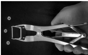
Hernia clamps: Cheap, designed to cause necrosis and scarring. Typically two pieces of wood are tightened across the hernia in an attempt to scarify the area. Complications include sepsis (gangrene) and evisceration.
Elastrator rings: use in foals around 3 months old with reducible hernias < 8cm in diameter. An applicator is used to apply the elastrator rings (usually 3 bands placed). **If the foal appears in severe pain after application, intestinal contents may be stuck in the rings and should be removed and evaluated.**
Hernia belts may or may not work better than mother nature; research is limited to non-existent. Many horse owners like hernia belts.
Herniorrhaphy: Surgery indicated if hernia is too large to attempt nonsurgical treatment, becomes incarcerated, or fails to resolve spontaneously by 6-12 months of age. Surgical repair can be done either closed (keep the hernia sac and peritoneum closed) or open (open the hernia sac and open the peritoneal cavity). Closed repair is frequently performed in horses. A fusiform incision is made around the hernia and the hernial sac dissected free of the skin (which is removed). The sac is pushed into the abdomen and the hernia sides closed over it.
Open repair is advised for bovine hernias since infection is often the cause of the hernia. After dissection to the abdominal wall, a stab incision is made into the abdominal cavity and then the hernia sac opened carefully, following the hernial ring. The stab incision should be made to one side to avoid the umbilical vessels that traverse cranially and caudally. These vessels should not be patent at this stage but may contain pus!
Body wall closure should be done with relatively large, long-lasting suture in a simple continuous pattern. We use #2 PDS for most weanlings. Babies like to bounce. Bouncing is hard on the incision. PDS is the longest lasting absorbable suture we have. It maintains strength for about 45 days. Continuous patterns have been shown to be more secure under tension than interrupted patterns. Reference this study, which is one of Dr. Malone’s all time favorites:
Comparison of incisional bursting strength of simple continuous and inverted cruciate suture patterns in the equine linea alba. Magee AA1, Galuppo LD.
OBJECTIVE: To determine the bursting strength of ventral median abdominal incisions closed by either simple continuous or inverted cruciate suture patterns.
METHODS: A 25 cm ventral median incision was made through the linea alba and a 200 L polyurethane bladder was placed within the abdomen. Either a simple continuous or an inverted cruciate pattern using 3 polyglactin 910 with a bite size and suture interval of 1.5 cm was used to close linea incisions. Closure time was recorded for each pattern. The bladder was inflated with air at 40 L/min, and the pressure at body wall failure recorded. The length of suture used for wound closure and the wound failure modes were recorded. Deviation from the linea (cm), total suture length (cm), suture length to wound length ratio (SL:WL), closure time (min), bursting pressure (mm Hg), and failure modes were compared between groups using Welch-Aspin t-tests. The effects of independent subject variables were assessed for possible effects on bursting strength using analysis of covariance.
RESULTS: Mean bursting pressure was significantly greater for the simple continuous pattern than for the inverted cruciate pattern (P = .01). Significantly less suture material (P = .0002) was required with the continuous pattern than with the inverted cruciate pattern. Mean closure time, SL:WL, deviation from the linea, and failure modes were not significantly different between groups. No significant effects were noted for independent variables in both groups on bursting strength.
CONCLUSIONS: In this model, a simple continuous closure pattern for ventral median abdominal incisions was stronger than an inverted cruciate pattern. A simple continuous pattern leaves less foreign material in the wound, which may be of benefit in reducing incisional complications.
CLINICAL RELEVANCE: Use of a continuous closure pattern for the linea alba may offer greater wound security during episodes of increased intra-abdominal pressure in horses.
We do recommend continuous patterns for all 3 layers of the body wall closure: body wall, subcutaneous tissue, skin.
Post operative care
Skin suture removal is at 14 days.
Restricted exercise/turnout for 1-2 months due to the slow healing of the body wall, the rapid loss of strength of most suture, and the likelihood that babies will still bounce. It is hard to charge for that second hernia repair if you didn’t advise the client on how to prevent it.
Complications
Many postoperative complications are similar across species for abdominal surgery and include infection, peritonitis and hernia recurrence.
Hernia recurrence
The body wall is fascia and is poorly vascular. In the adult horse, it takes approximately 60 days for the hernia repair to have moderate strength. No absorbable suture lasts that long so restricted exercise is important to prevent hernia recurrence.
Richter’s and entrapped hernias
If the intestines are stuck, the animal will usually show signs of colic, at least initially. Eventually the animal may become depressed. In a normal (reducible) hernia, the intestines can be shoved back into the belly; if the intestines are entrapped, the hernia is not reducible. The hernia area may be painful on palpation. The area may be warmer or colder than surrounding areas.
With a Richter hernia, only part of the intestinal wall is entrapped vs the full width of the intestine being entrapped. This part of the intestinal wall can become devitalized and leak, resulting in an intestinal fistula.
Key Takeaways
- Umbilical hernias are often associated with umbilical infections in calves.
- Pigs and foals just get hernias (not always infections) and these are likely hereditary. We still do surgery on foals but owners should be advised about inheritance risk. We try to avoid surgery in show pigs.
- Use fusiform incisions, enter abdominal cavity to the side (not cranial or caudal).
- Foals with umbilical infections are sick. Since the diagnosis occurs fairly early, these animals do respond to antibiotic therapy.
- The body wall heals really slowly. Early exercise will increase the risk of hernia recurrence.
Resources
Management of umbilical disorders in the foal, In Practice, 2008- more details; nice reference for your files
Tissue Strength and Wound Morphology of the Equine Linea Alba After Ventral Median Celiotomy, Vet Surg 2000- evidence regarding how long the body wall takes to heal after surgery
Video: bandaging a hernia to keep it reduced
Video: field anesthesia for hernia repair – designed to give you an mental image of the procedure
Video : Simple hernia repair in a horse (or dog, pig, calf, etc)- more specifics for those that want them
Video: protecting the intestines during body wall closure – cool technique that applies to all species; youtube link
