FA GI Topics
How to – Rectal prolapse submucosal resection
Indications
Submucosal resection is indicated when a prolapse is too damaged to be replaced but the damage is restricted to the mucosal layer, when the prolapse cannot be effectively reduced with osmotic agents only, or when the prolapse has recurred. A resection decreases the size of the tissue and minimizes the amount of tissue that can prolapse.
Relevant anatomy
The exposed tissue is mucosa.
Preoperative management
Food restrictions: NA
NSAIDs/analgesics: NSAIDs and an epidural are indicated.
Antibiotics: Preoperative antibiotics are indicated.
Tetanus prophylaxis is recommended for small ruminants.
Local blocks: Epidural. Lidocaine may also be infused into the rectum.
Position/preparation: Standing procedure if possible; otherwise sternal recumbency. The tail should be tied or taped away from the surgical field.
Surgery Supplies:
- Scalpel
- Needle holders
- Hemostats
- Thumb forceps
- Suture – 2-0 or 3-0 absorbable, taper needle
- Spinal needles to hold the rectum out while suturing
- Syringe case (rectal lumen diameter)
Surgical procedure
- Insert two 18ga 6″ spinal needles at right angles to each other, through external anal sphincter and healthy mucosa to keep the rectum prolapsed during dissection. Use older (duller) spinal needles. A syringe case is useful to keep the lumen identified and open.
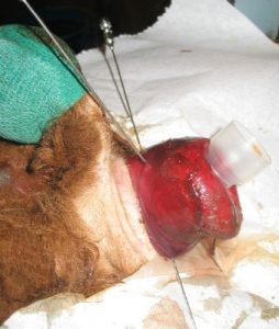
- Create two 360o incisions to the depth of the submucosal layer. The first should be as close to the anus as possible, the second as distal as possible.
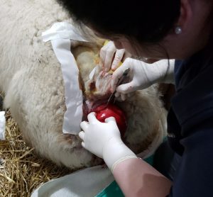
- Connect the two 360o circles with a vertical incision to the same depth
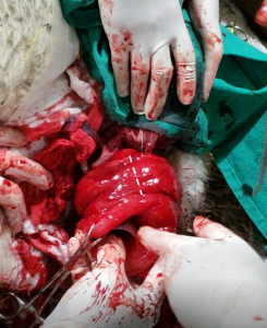
Starting at the connecting line, dissect off the mucosa to the submucosal layer. Continue around the full circumference.
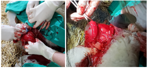
Suture the two 360o incisions back together using a continuous pattern using 2-0 or 3-0 absorbable suture material. Tie a knot midway to avoid a purse string effect.
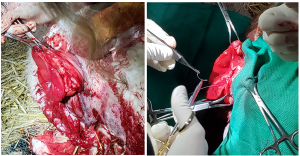
- Remove any spinal needles and syringe case.
Postoperative care
- Use a purse string suture around the rectum to keep the tissue in place
- tie to the side and leave in place for one week
- ensure opening is large enough for pellets to be evacuated
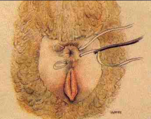
- Maintain soft feces using a legume diet and laxatives
- Continue NSAIDS for at least 3 days
- Epidural or lidocaine infusion if straining continues
- Tetanus booster for horses and small ruminants
- Horses may need digital removal of feces
Complications
This technique has fewer related complications than amputation but prolapse recurrence is possible.
Videos
Resources
Rectal Prolapse. Vet Clin NA FA 24 (2008) 403–408
the suture runs in and out in a circular pattern, so when pulled tight, it cinches off the center area like a purse string

