LA Respiratory Issues
Paranasal sinus disease
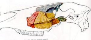
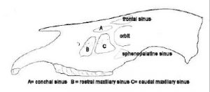
Large animal species have complex sinus anatomy, supposedly related to decreasing the weight of the head but mostly serving to create pockets of infection. The equine frontal and maxillary sinuses drain through the nasomaxillary opening in the rostral maxillary sinus.
Common paranasal sinus disorders with surgical therapies:
Primary sinusitis
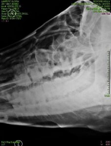
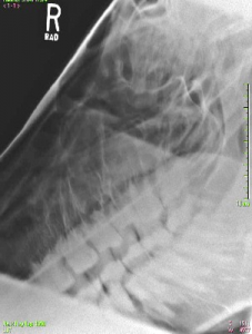 Sinusitis results from infections and when the nasomaxillary opening is obstructed and drainage is impaired. CT and Radiographs are useful to identify fluid in the sinus(es). Note two separate fluid lines in the radiograph to the left. Surgical treatment involves flushing the sinus and/or creating drainage into the middle nasal meatus. Warning: Streptococcus equi should be considered as a possible cause.
Clinical signs – unilateral or bilateral purulent nasal discharge; occasionally decreased airflow and no discharge
Traumatic sinusitis
Sinus trauma, including dehorning trauma, can lead to sinusitis. Drainage can be problematic if the trauma affects the frontal sinus. The frontal sinus needs to drain into the maxillary sinus before it can drain out the nasal passages. Blockage along the way can occur due to other consequences of trauma or to inspissated pus. Drainage may be through sinus trephination, through enlargement of the nasomaxillary openings or creation of new openings into the nasal passageway.
Clinical signs: unilateral or bilateral purulent nasal discharge; occasionally decreased airflow and no discharge
Tooth root infections
 The maxillary sinus of the horse is divided into rostral and caudal compartments by a septum. In adult horses, the roots of teeth 109 and 209 (sometimes 108, 208) enter the rostral maxillary sinus and the roots of teeth 110, 111, 210, 211 enter the caudal maxillary sinus. These roots of these teeth can become infected and lead to secondary infection of the maxillary sinus. Tooth root infection should be suspected with unilateral sinusitis. While radiographs are useful to identify sinusitis, CT (computed tomography) is the gold standard for determining if a tooth (teeth) are infected. If the teeth are NOT in the sinus (more rostral), they can still get root infections but now we get draining tracts and fistula.
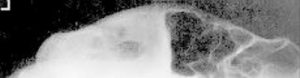
In the radiograph to the left, diastemata (gaps) are visible between multiple teeth and teeth are missing or abnormal in shape.
Clinical signs: unilateral purulent nasal discharge or orocutaneous fistula, diastema on oral exam
Sinus cysts 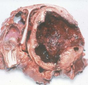
Sinus cysts are similar to conchal cysts in development, clinical signs, and therapy. Radiographs and CT examinations are useful for diagnosis.
Clinical signs: decreased airflow through one or both nostrils
Ethmoid hematomas
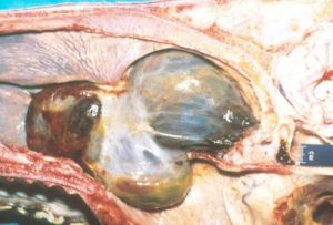 Definition of ethmoid hematoma: Well-encapsulated masses originating in or around the ethmoid labyrinth or paranasal sinuses.
Etiology: The cause of ethmoid hematomas is unknown but hemorrhage occurs in the submucosa of the turbinate or sinus, causing the mucosa to stretch and thicken, forming the capsule of the hematoma. Affected horses are usually middle aged. The ethmoid hematomas can be unilateral (most common) or bilateral.
Clinical signs: Low grade epistaxis (nose bleed) +/- nasal discharge. Lesions are almost always unilateral.
Diagnosis: Endoscopy combined with radiographs or CT is useful for diagnosis and evaluation of size and location.
Treatment: Treatment options include laser therapy, formalin injections and/or surgery (sinus flap to remove it). Recurrence is common. This is a horse thing. No one is quite sure why these don’t just resolve like most hematomas.
Clinical signs: Intermittent unilateral epistaxis +/- nasal discharge.
Sinus tumors 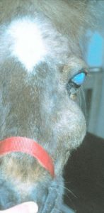
Sinus tumors are uncommon but include squamous cell carcinoma and tumors of dental origin. Clinical signs are related to bony remodeling, tissue necrosis and impairment of nasal discharge. CT is a gold standard for diagnosis. Sinus flaps may be needed for diagnosis and therapy. Tumors are often advanced by the time of diagnosis.
Clinical signs: unilateral purulent nasal discharge or decreased airflow
Challenge Yourself
Level A
Dental disorders are common. Work your way through the following exercise to review the topic.
Resources
Surgery of the paranasal sinuses of horses, Equine Vet Educ. 2024;36:206–216.- not really field surgery but shows nerve blocks and the procedure
Disorders of the paranasal sinuses, Tremaine and Freeman – great pictures and explanations
Paranasal sinus disease, 2011 Compendium- another great resource for common diseases
Respiratory distress in the adult and foal, VCNA (2021) 37:311-325
How to perform a minimally invasive sinus flush, 2008 AAEP – very handy skill if doing equine practice
Sinusitis in horses, ACVS -short and sweet with a surgery perspective
Manual of Clinical Procedures in the Horse
Frontal sinusitis in adult beef bulls, JAVMA 2019
Anatomy and Diagnostic imaging of the equine paranasal sinuses – Apple ibooks from UGA
