Bovine Lameness and Podiatry
Sole ulcers
G Cramer
What is it?
Sole ulceration is one of the three most common causes of lameness affecting beef and dairy cattle. Sole ulcers occur beneath the flexor tuberosity of P3 (third phalanx). These occur most commonly in the outside digit in rear legs and are associated with varying degrees of changes in weight bearing.
How to recognize it?
Sole ulcers are recognized by the presence of severe hemorrhage or protrusion of the corium at the typical sole ulcer site. Hemorrhage or bruising may be evident. Hoof tester pressure in the area will result in a withdrawal response due to pain. Severe hemorrhages with an associated pain withdrawal reflex upon pressure with hoof testers should be considered early sole ulcers and treated accordingly.

Pathogenesis
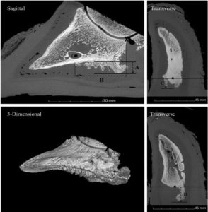
Sole ulcers are due to continuous pressure by the flexor tuberosity of P3 on the corium. This pressure is caused by mechanical or metabolic alterations in the supporting structures of the hoof (P3 is allowed to drop).

This pressure initially leads to the corium leaking blood into keratinocytes at the dermal-epidermal interface. Over time this pressure from P3 leads to the destruction of keratinocytes and the interruption of horn growth resulting in the corium protruding through the horn defect.
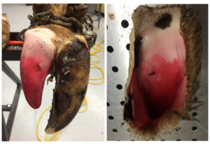
The resultant inflammation also results in long term structural changes to P3 and the corium.
How do you prevent a sole ulcer?
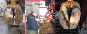
The prevention of sole ulcers consists of ensuring adequate lying time, minimizing negative energy balance (making sure they have adequate body condition) and following an appropriate hoof trimming schedule.

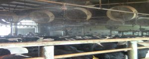
To ensure a lying time of 12-14 hours, cows should not be away from their pen for more than 3-4 hours. In addition, forced standing time should be kept to a minimum and effective cooling strategies to reduce the impact of heat stress on standing time should be implemented.
It is important to make sure first lactation animals are adjusted to adult cow housing at least 60 days prior to calving. .Finally the strategic use of an appropriately timed and correctly performed hoof trimming should be a key component of a prevention program.
How do you treat a sole ulcer?
Sole ulceration results in chronic changes and is a painful condition. Appropriate early treatment is critical to successful resolution of symptoms and to minimize the impact of long term changes. NSAIDs or other analgesics are indicated.
The treatment of sole ulcers involves the removal of all loose horn around the corium. This removal should be done delicately, with great care taken to minimize further damage to the corium.
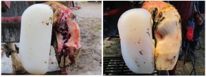
Once loose horn has been removed around the lesion, pressure on the lesion should be reduced to maximize the speed of horn growth. The reduction of pressure on the lesion is is achieved by the removal of horn around the periphery of the lesion and by application of a properly sized hoof block to transfer weight to the sound digit.
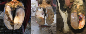
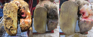
Lameness should improve gradually but can take 2-6 weeks to resolve. Cows with sole ulcers should be rechecked in 3-6 weeks to assess healing, and to either remove or reposition the block if necessary.
Key Takeaways
Sole ulcers develop as the flexor tuberosity of P3 pushes on the sole from inside the foot. This occurs due to loss of he supporting structures in the foot. Treatment includes removing loose horn and placing a block on the opposite digit.
Prevention is key. Cows should have comfortable areas in which to lie down, be on a regular trimming schedule, and be on a good nutritional plane.
Resources
Lameness originating in the hoof of cattle, Cramer and Solano, merckvetmanual.com
Implications for lameness control in cattle -Vet Clin Food Anim 41 (2025) 395–406
The DairyLand Initiative -good pictures
dairyknow.umn.edu – lots more about foot issues and risk factors

