Equine Oral, Esophageal and Rectal disorders
Esophageal stricture
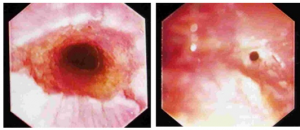
Esophageal strictures may be congenital or occur secondary to surgery; however, most occur secondary to trauma from choke. If the choke leads to erosions in the mucosa, fibrosis may lead to stricture. This is particularly a problem with circumferential erosions. As the esophagus heals, it contracts in diameter for the first 15 days, becoming narrower and narrower. Between days 30 and 60, the lumen diameter actually increases. The risk of recurrrent choke is very high in the first 60 days. The diameter will not usually improve after 60 days.

Acute cases may resolve with nonsurgical management. Horses are treated with antibiotics (choke can lead to aspiration pneumonia), soft feed, and NSAIDs. Bougeniage or balloon dilation has been helpful. Topical or systemic corticosteroids can be used to minimize scarring after infection is controlled. Surgery should be postponed until at least 60 days to allow full diameter to be achieved. Surgery is uncommon; referral is indicated.
Resources
Oesophageal disorders in horses: Retrospective study of 39 cases, EVE 2018, Vol.30(2), pp.94-99
Conservative treatment of acquired oesophageal strictures by endoscopic-guided balloon dilation in two horses, EVE 2017, Vol.29(5), pp.259-265
Diagnosing disorders of the equine oesophagus, EVE 2015, Vol.27(6), pp.291-294

