Equine Lameness
Distal limb bones review
Really cool 3d distal limb model
Fetlock and Digit Set Bone Box- Dr. Cox
you can check in with anatomy or a year 1 buddy and see if you can review using a bone box!
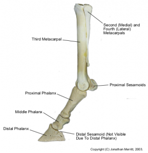
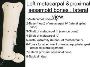
Distal cannon bone (metapodium = metacarpal or metatarsus)
- medial and lateral fossae provide attachment sites for the collateral ligaments.
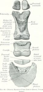
- The condyle is the distal articular surface of the cannon bone and P1 and P2. Note, however, that the distal cannon has a sagittal ridge while the condyles on the distal ends of P1 and 2 are saddle shaped.
Proximal phalanx (P1)
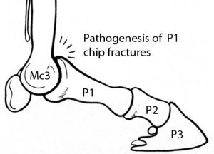
Note the sagittal groove on the proximal end of P1. This groove interdigitates with the sagittal ridge on the end of the cannon bone. A “screwdriver” fracture starts at this groove and runs longitudinally to split P1 into 2 pieces. The screw driver analogy pictures the sagittal ridge of the cannon bone as the blade of a screwdriver and the proximal end of P1 as the head of a screw. According to JR Rooney, these fractures are caused by rotary motion and acceleration. Consider a screw in oak being unable to twist as rapidly as the screwdriver above it resulting in fracture of the screw.
Hold P1 and the cannon bone together as they articulate. Note that the dorsal proximal edge will butt up against the dorsal surface of the cannon bone when the fetlock joint is over extended as happens during extreme downward translation of the fetlock. This is another consequence of excessive downward translation of the fetlock region. Previously mentioned were fractures of the sesamoid bones and bowed tendons.
On the palmar/plantar surface find the triangular area where the middle distal sesamoidean ligaments are inserted. On the distal end of P1 and P2 note the shallow indentation of the articular surface to form a saddle shaped surface as described by radiologists. On the palmar surface of the distal end of P1 are facets for attachment of the SDF tendon.
Proximal sesamoids
The proximal sesamoid bones are what make the fetlock the widest part of the distal limb (other than the hoof). Aside from their articular surfaces, the rest of the sesamoids are covered with a variety of tendons and ligaments. Therefore, even after removal of the skin and loose subcutaneous tissue, the sesamoids are hidden from view by their many attachments.
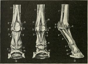
- The fibrocartilagenous intersesamoidean ligament binds the axial surfaces together to form a groove on the palmar surface for the digital flexor tendons. On the articular surface is a narrow groove that interdigitates with the sagittal ridge of the distal cannon.
- Note that the articular surface of the proximal sesamoids is the only smooth surface
- Abaxial surfaces have a roughened depression for insertion of the suspensory ligament that covers the entire abaxial surface.
- Basal surface is covered by the attachments of the distal sesamoidean ligaments.
Middle phalanx (P2)
This is a short very compact bone. Proximal phalanx (P1) is twice as long as the middle phalanx (P2). Note the strong proximal palmar (planter) edge. This area is strengthened for insertion of the superficial distal sesamoidean ligament and the SDF tendon. Not seen is the attached complementary fibrocartilaginous ridge of P2 which is part of the insertions. On the sides of the ends of P1 and P2 there are depressions for attachment of the collateral ligaments of the PIP and DIP joints respectively.
Distal phalanx = P3 = coffin bone.
The extensor process on the coronary border is the place of insertion of the main extensor tendon. Just behind the extensor process note the depression on the lateral side of the coffin bone for attachment of the collateral ligaments of the coffin joint.
The palmar/plantar extensions of P3 are often referred to as the wings of P3 by radiologists. This is the site for attachment of the collateral cartilages of the coffin bone. Unfortunately these cartilages as often referred to as the hoof cartilages but this is a misleading term because the cartilages are attached to P3 and not the hoof. Instead, the collateral cartilages are inside the hoof but their upper parts are above the coronet where they can be palpated easily in the live horse. The collateral cartilages are attached to the wings of P3 just behind the depressions for the collateral ligaments and extend into the heel beyond the coffin bone
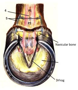
Distal sesamoid bone = Navicular bone
This bone gets the popular name from its boat shape. Note that the distal edge is rounded like the bottom of a boat, the proximal surface is straight like the deck of a boat. Note the foramena on the distal edge. These are probably for blood vessels, but if the blood vessels recede, the foramena may contain invaginations from the distal palmar pouch of the coffin joint. The other surfaces are the flexor surface adjacent to the navicular bursa and the articular surface which forms part of the wall of the coffin joint = DIP joint.
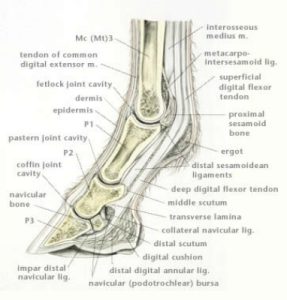
Split limbs study
Find the following: DDFT, SDFT, pastern (PIP) joint, straight sesamoidean ligament, navicular bursa, coffin (DIP) joint pouches (dorsal, proximal and distal palmar), digital cushion and distal sesamoidean impar ligament.
Summary of synonyms
- Interosseous tendon = suspensory ligament (suspends the fetlock and sesamoids)
- Proximal phalanx = long pastern bone = P1
- Middle phalanx = short pastern bone = P2
- Distal phalanx = coffin bone = P3
- Superficial distal sesamoidean ligament = straight sesamoidean ligament (Z) [unpaired]
- Middle distal sesamoidean ligament = oblique sesamoidean ligament (Y) [paired]
- Deep distal sesamoidean ligament = cruciate sesamoidean ligament (X) [paired]
- Collateral cartilage of P3 = hoof cartilage = ungual cartilage
- Proximal interphalangeal joint = PIP joint = pastern joint
- Distal interphalangeal joint = DIP joint = coffin joint
- Podotrochlea bursa = navicular bursa
- (Podotrochleosis = navicular disease)
inflammation and swelling of tendons that makes them have a bowed appearance (curved)
