13.4 Male reproductive anatomy
Some of the organs involved with male reproduction are diagrammed below.
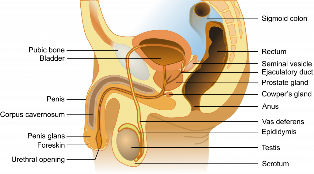
Male reproductive anatomy involves the organs and glands that produce sperm, create semen to transport sperm, and conduct this liquid semen out of the body. Semen production involves the work of accessory glands, each responsible for the production of one or more key ingredients of semen. Male anatomical structures can be broken down into the following:
Sperm production
- Testis: males typically have two testes (also called testicles), which, in humans, descend from the abdomen during fetal development and are enclosed outside the abdomen in the scrotum. Each testis houses many tube-like structures (the seminiferous tubules) in which sperm are made. Specialized cells (the Leydig cells) in the testes produce testosterone.
- Scrotum: a pouch of skin that holds the testes that contracts or expands to adjust the distance the testes are from the body to regulate their temperature.
- Seminiferous tubules: these structures within the testes are the actual sites of sperm production (discussed further below)
- Epididymis: this rubbery device sits astride the testis. Sperm mature here and are stored prior to ejaculation (when sperm-bearing semen leaves the body, typically during orgasm)
Semen production
- Seminal vesicles: these two glands produce an alkaline (basic) fluid that can neutralize the acidity of the vagina. This fluid contains fructose and other nutrients to provide energy for the sperm.
- Bulbourethral (or Cowper’s) glands: these two glands provide a mucus-rich alkaline fluid that lubricates the inside of the urethra to allow for easier passage of sperm and neutralizes the urethra (urine residue is acidic). Some of this fluid exits the penis prior to ejaculation (this pre-ejaculate fluid can also contain sperm). The remainder of the fluid combines with the semen ejaculate.
- Prostate gland: this organ wraps around the urethra and provides muscular contractions that help propel semen during ejaculation and block urine flow from the bladder during ejaculation. It also provides fluid in the ejaculate that contains enzymes and zinc that aid in sperm motility.
Sperm/semen transport:
- Ductus (or vas) deferens: this pair of muscle-lined tubes carry sperm from the epididymis of each testis into the abdominal cavity where they loop over the bladder and join with the ducts from the seminal vesicles to form the ejaculatory ducts. The muscles that line the ductus deferens contract to propel semen during ejaculation.
- Ejaculatory ducts: these ducts are formed by the joining of the vas deference with the duct from the seminal vesicle. Each ejaculatory duct empties into the urethra.
- Penis: the organ that encircles the urethra as the urethra exits the abdomen. This organ changes from flaccid (soft and limp) to erect (rigid and standing away from the body) during sexual arousal or spontaneously. In uncircumcised men the penis has a fold of skin called a foreskin that during the flaccid state, covers the head of the penis, and during the erect state retracts behind the glans (or head) of the penis.
- Urethra: the tube that runs from the bladder through the penis through which urine and semen exit the body.
