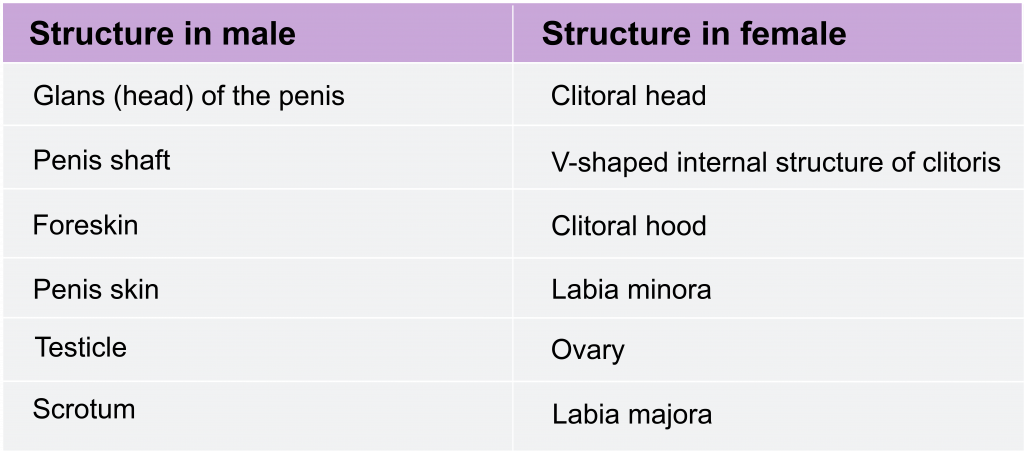
13.3 Commonalities between male and female reproductive anatomy
During embryonic development the male and female fetus are indistinguishable before about 10 weeks of pregnancy. Fetal tissues begin in an undifferentiated state, and based on genetic signals and the interuterine environment the reproductive organs usually differentiate into structures typical of males and females (for illustrations see chapter 8.6). This means that for most of the reproductive parts there is an analogous part in the other sex that arose from the same original tissues. For example, testes and ovaries develop from the same tissue – originally located in the abdomen. In males the testes move down and outside the abdomen as they develop; in female they remain internal. Some structures (such as the oviducts) have a structure that was common in early development, but completely or partially disappears in later development; other structures (such as the uterus) have analogues that are very subtle structures in the male. See the following table for a list of analogous structures in male and female anatomy.
Commonalities between male and female reproductive signaling
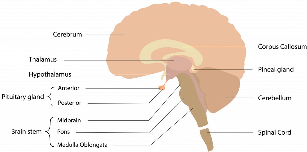
Much of the reproductive physiology we will address is regulated by hormonal signals that arise in the brain and much of this signaling is shared between males and females.
Within the brain is a region called the hypothalamus (see figure 2). This portion of the brain sends signals to the pituitary gland located beneath it. In particular, the hypothalamus sends a hormonal signal called gonadotropin-releasing hormone (GRH) to the pituitary gland. In response to the GRH signal, the pituitary gland releases two hormones that circulate in the blood: luteinizing hormone (LH) and follicle stimulating hormone (FSH). These hormones travel throughout the body, triggering further hormone releases and physiological changes (discussed further below). There are feedback loops that tightly regulate the levels of circulating hormones. In addition to GRH, LH, and FSH, the hormones testosterone, estrogen and progesterone are important in reproductive signaling. While we will focus on the effects of testosterone in males and estrogen and progesterone in females, all of these hormones are present and important in both males and females.
In the following pages the reproductive anatomy and physiology of male and female reproduction are described. We begin with reproductive anatomy, which describes the organs and tissues involved in reproduction, and then go on to describe the physiology, or how these structures function together and respond to hormonal signals
