7 Hepatic lipidosis
Pathophysiology
Cats, camelids, and cows tend to get fatty liver when they are off feed or in a negative energy balance. Miniature horses can do this too. Pregnancy toxemia in sheep is related. Obese animals are particularly at risk.
The disorder is very common in cats and is responsible for almost half of the cases of feline liver disease. This is the biggest reason why cats should not be allowed to become overweight. A sick cat tends not to eat and this leads to fat mobilization and hepatic lipidosis.
In cattle, this generally occurs in early lactation as the cows cannot eat enough to meet their metabolic demand and try to mobilize fat stores. In animals affected with hepatic lipidosis, metabolic derangements and liver dysfunction occur due to increased mobilization of fatty acids.
- Hyperlipemia occurs when triglycerides are not removed from the blood as fast as they are produced.
- Lipidosis occurs when triglyceride synthesis exceeds formation and release triglycerides into circulation
Normal physiology
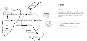
With lipolysis, serum free fatty acid (FFA) concentration increases. These FFAs are absorbed by liver cells and metabolized. Some go through carnitine dependent oxidation and produce energy in the TCA cycle or form ketone bodies. In this pathway, extra FFA are made into triglycerides and stored in hepatocellular vacuoles. They get secreted into the circulation after incorporation into very low density lipoproteins (VLDLs).
Lipidosis
With lipidosis, several things could alter the pathway
- These animals may have insufficient protein to form VLDLs which are important for removal of excessive triglycerides
- Relative amounts of carnitine may be too low to transport fatty acids thru the liver
- Insulin resistance. Normally fat breakdown occurs in response to insulin. Poor insulin function may lead to continued lipolysis in the face of energy restriction.
Current thinking is protein deficiency combined with insulin issues.
Diagnosis
Animals become anorexic (lose appetite), lose weight, lose muscle mass, and become icteric. Cattle drop in milk production. The liver enlarges as it fills with fat.
Serum liver enzymes increase due to hepatocellular liver damage. Cholestasis occurs as the bile ducts get filled with lipids and collapse.
Liver ultrasound and/or biopsy is diagnostic in cattle.
Therapy
Therapy is focused on resolving the energy gap and supporting the liver.
Treatment in cattle includes providing iv glucose, insulin, vitamins and potentially steroids. Rumen transfaunation may also be useful.
Cats often need to be fed via a surgically placed feeding tube for several weeks. Fluids and anti-nausea drugs are often indicated. Potassium and glucose may need supplementation.
Resources – cattle
Hepatic lipidosis in ruminants, VCNA, 2023 – biochemistry and more biochemistry
Hepatic lipidosis in cattle, Purdue, 2003
Clin path evaluation of hepatic lipidosis in cattle, JVIM, 2008
Prediction and diagnosis of fatty liver in dairy cattle, SMJGH, 2017
Fatty liver disease in cattle -Merck
Resources – cats
Hepatic lipidosis in cats – Blue Pearl
Resources – Hyperlipemia in horses
Hyperlipemia in donkeys, The donkey sanctuary
Equine hyperlipidemias, VCNA 2011
Hyperlipemia mini horses and donkeys, JVIM 1994
Severe hypertriglyceridemia in clinically ill horses, EVJ 2003
breakdown of fats
